. 2009 Nov 10;106(45):19056-60.
doi: 10.1073/pnas.0904805106. Epub 2009 Oct 26.

Complex embryos displaying bilaterian characters from Precambrian Doushantuo phosphate deposits, Weng'an, Guizhou, China

Affiliations

Complex embryos displaying bilaterian characters from Precambrian Doushantuo phosphate deposits, Weng'an, Guizhou, China

Jun-Yuan Chen et al. Proc Natl Acad Sci U S A. .

Abstract

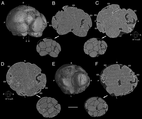
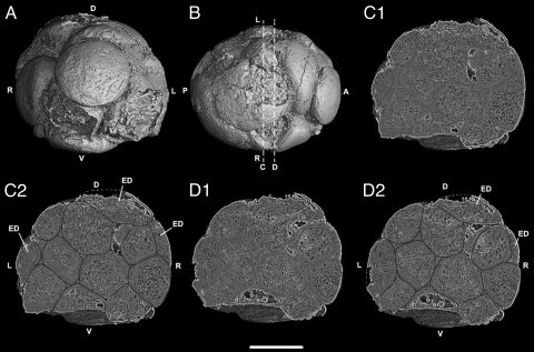
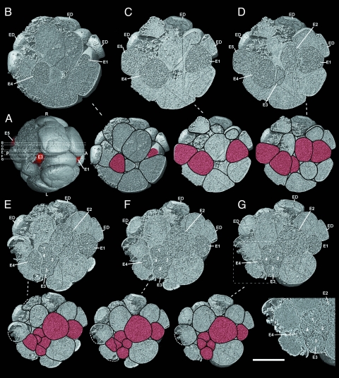
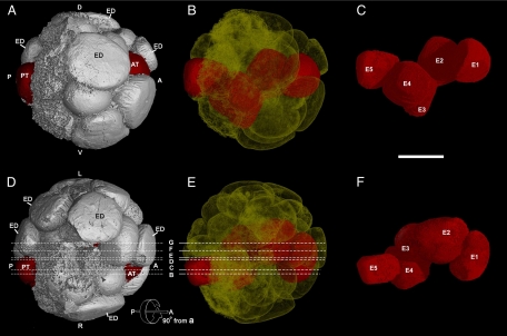
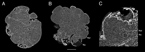
Three-dimensionally preserved embryos from the Precambrian Ediacaran Doushantuo Formation, Weng'an, Guizhou, southern China, have attracted great attention as the oldest fossil evidence yet found for multicellular animal life on Earth. Many embryos are early cleavage embryos and most of them yield a limited phylogenetic signal. Here we report the discovery of two Doushantuo embryos that are three-dimensionally preserved and complex. Imaging techniques using propagation phase-contrast based synchrotron radiation microtomography (PPC-SR-microCT) reveal that the organization of cells demonstrates several bilaterian features, including the formation of anterior-posterior, dorso-ventral, and right-left polarities, and cell differentiation. Unexpectedly, our observations show a noticeable difference in organization patterns between the embryos, suggesting that they represent two distinct taxa. These embryos provide further evidence for the presence of bilaterian animals in the Doushantuo biota. Furthermore, these bilaterians had already diverged into distantly related groups at least 40 million years before the Cambrian radiation, indicating that the last common ancestor of the bilaterians lived much earlier than is usually thought.


Conflict of interest statement

The authors declare no conflict of interest.

Figures

Fig. 1.
Fig. 1.
Precambrian postgastrular embryo (4F10) from the Weng'an Phosphate Member of the Doushantou Formation showing external view from right side (A) and ventral side (E), and digital internal sections (B, C, D, and F) and the same sections (B, C, D, and F) with drawings of cell boundaries (in reduced size) along the a-p axis. (A) External view from right side along the anterior (right) and posterior (left) axis with lines C and D showing the locations of the internal digital sections seen in Fig. 2. (B) Digital internal section and the same section with drawing of cell boundaries at reduced size cut half way into the embryo from the right surface (Fig. 1A), showing maximal dorso-ventral asymmetry and ectodermal cells. (C) Digital internal section and the same section with drawing of cell boundaries at reduced size along the same (a–p) axis as Fig. 1B with 30° rotation top down away from the observer showing transition from asymmetry (Fig. 1B) to bilateral subsymmetry (Fig. 1D). (D) After 90° rotation from Fig. 1B, internal section and the same section with drawing of cell boundaries at reduced size showing the plane of left-right bilateral subsymmetry. (E) External digital view from the ventral side, revealing exposed, putative endodermal cells. (F) Internal section and the same section with drawing of cell boundaries at reduced size cut inward 23% from ventral surface (Fig. 1E), showing left-right, bilateral subsymmetry of the massive internal cells. (Scale bar, 250 μm.) Key: A, anterior; P, posterior; D, dorsal; V, ventral; R, right side; L, left side; ED, ectodermal cell.
Fig. 2.
Fig. 2.
Precambrian postgastrular embryo (4F10) from the Weng'an Phosphate Member of the Doushantou Formation showing external view from apical pole (A) and dorsal side (B) with two digital internal sections (C1, D1) and the same sections with drawings of cell boundaries (C2, D2), at locations indicated at lines C and D (Fig. 1A, Fig. 2B). (Scale bar, 250 μm.) Key: A, anterior; P, posterior; D, dorsal; V, ventral; R, right side; L, left side; ED, ectodermal cell.
Fig. 3.
Fig. 3.
Precambrian postgastrular embryo (4F4) from the Weng'an Phosphate Member of the Doushantou Formation as serial digital sections that cut in at successively deeper layers from the right surface, indicated by the lines B–G both in Fig. 3A and Fig. 4 D and E, and the same sections with drawings of cell boundaries. (A) Digital external view from ventral side of the embryo 4F4, showing: E5, E3, and E1 cells (in red) and the positions of digital internal sections in Fig. 3 B–G indicated by lines B–G. (B) Internal section and the same section with drawing of cell boundaries at reduced size cut in 29% from right surface, showing principal endodermal cells E4 and E1 sectioned near the right margin of the endodermal cord. (C) 35% from right surface, showing endodermal cells E5, E4, and E1. (D) 43% from right surface. (E) 46% from right surface. (F) 51% from right surface. (G) 53% from right surface, showing most parts of the endodermal cord, close to the middle plane of the embryo, and cluster of four cells, apparently the daughter cells resulting from mitosis of E3 (enlarged in lower right panel). Key: A, anterior; P, posterior; D, dorsal; V, ventral; L, left side; R, right side; and ED, ectodermal cell. In reduced sections with cell boundaries drawn endodermal cells are in red. (Scale bar: 250 μm in digital sections, 375 μm in the same section with drawing of cell boundaries, 181 μm in the lower right panel.)
Fig. 4.
Fig. 4.
Precambrian postgastrular embryo (4F4) from the Weng'an Phosphate Member of the Doushantou Formation. (A) Digital external view from right side along the anterior (right: A) and posterior (left: P) axis, showing: anterior (AT) and posterior (PT) terminations (in red) of the putative endodermal cord of cells, and presumed ectodermal cells on dorsal surface. (B) Transparent view from right side, showing the putative endodermal cord of cells (in red) that has a shallow S shape dorso-ventrally, and extends through the embryo from anterior to posterior ends. (C) Digital view of the putative endodermal cord seen laterally from the right side with its constituent cells coded as E1–E5. (D) Digital external view from dorsal side of the embryo, showing: the cells at the anterior (AT) and posterior (PT) terminations of the putative endodermal cord (in red); the lines designated B–G represent the locations of digital sections shown in Fig. 3 B–G. (E) Digital transparent view from dorsal surface, showing the putative endodermal cord (red), curved into a shallow S shape laterally; the lines B-G representing locations of the section shown in Fig. 3 B–G. (F) Digital view of the endodermal cord from dorsal side and its constituent cells coded as E1–E5. (Scale bar, 250 μm.) The endodermal cells were isolated and colored with the segmentation program of VG studio Max 1.21. Key: A, anterior; P, posterior; AT, anterior termination; PT, posterior termination; D, dorsal; V, ventral; L, left side; R, right side.
Fig. 5.
Fig. 5.
Preservation of yolk granules and possible nucleus. (A) Internal section cut inward 39% from dorsal surface, showing detail of the yolk-rich, internal cells in embryo 4F10. (B) A digital thin slice (0.1-μm thick) of an internal section of embryo 4F4 46% from right surface (rotated, but same plane of section as in Fig. 3E), revealing the yolk-rich endodermal cells, as well as the location of a putative nucleus and nucleolus. (C) Internal section of Fig. 5B cell showing a close-up of the putative nucleus and nucleolus, as seen in Movie S3. Key: Nu, nucleus; Nul, nucleolus. (Scale bar, 142 μm in A, 156 μm in B, and 49 μm in C.)

References

    1. Condon D, et al. U-Pb ages from the Neoproterozoic Doushantuo Formation, China. Science. 2005;308:95–98. - PubMed
    1. Chen DF, Dong WQ, Zhu BQ, Chen XP. Pb-Pb ages of Neoproterozoic Doushantuo phosphorites in South China: Constraint on early metazoan evolution and glaciations events. Precambrian Res. 2004;132:123–132.
    1. Barfod GH, et al. New Lu-Hf and Pb-Pb constraints on the earliest animal fossils. Earth Planet Sci Lett. 2002;201:203–212.
    1. Hagadorn JW, et al. Cellular and subcellular structure of Neoproterozoic animal embryos. Science. 2006;314:291–294. - PubMed
    1. Xiao S, Zhang Z, Knoll AH. Three dimensional preservation of algae and animal embryos in a Neoproterozoic phosphorite. Nature. 1998;391:553–558.

Publication types