. 2009 Nov 10;106(45):18960-5.
doi: 10.1073/pnas.0906734106. Epub 2009 Oct 26.

LACTB is a filament-forming protein localized in mitochondria

Affiliations

LACTB is a filament-forming protein localized in mitochondria

Zydrune Polianskyte et al. Proc Natl Acad Sci U S A. .

Abstract

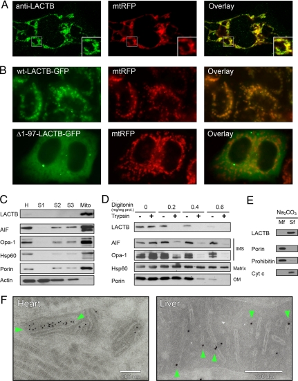
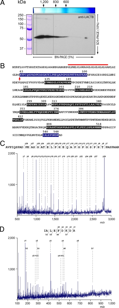
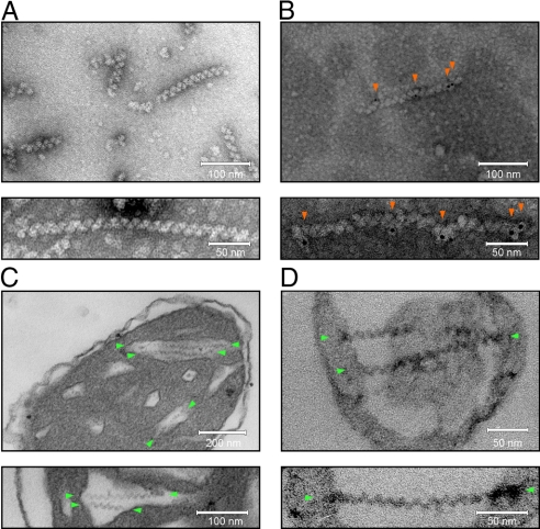
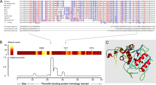
LACTB is a mammalian active-site serine protein that has evolved from a bacterial penicillin-binding protein. Penicillin-binding proteins are involved in the metabolism of peptidoglycan, the major bacterial cell wall constituent, implying that LACTB has been endowed with novel biochemical properties during eukaryote evolution. Here we demonstrate that LACTB is localized in the mitochondrial intermembrane space, where it is polymerized into stable filaments with a length extending more than a hundred nanometers. We infer that LACTB, through polymerization, promotes intramitochondrial membrane organization and micro-compartmentalization. These findings have implications for our understanding of mitochondrial evolution and function.


Conflict of interest statement

The authors declare no conflicts of interest.

Figures

Fig. 1.
Fig. 1.
Subcellular localization of LACTB in HeLa cells and rat tissues. (A and B) Fluorescence microscopy images of mtRFP. (A) Endogenous LACTB visualized with an anti-LACTB antibody and an Alexa 488-coupled secondary antibody. The region marked with a square is enlarged (Inset). (B) Transfection with plasmid constructs encoding WT LACTB or an N-terminally truncated LACTB fused to a C-terminal GFP (wt-LACTB-GFP, and Δ1–97-LACTB-GFP). (C) Immunoblotting of proteins from rat liver homogenate (H), cytosolic fractions (S1-S3), and mitochondrial fraction (Mito). (D) Immunoblotting of LACTB from rat liver mitochondria incubated with digitonin and trypsin compared with compartment-specific markers for intermembrane space (IMS), matrix, and outer membrane (OM). (E) Soluble mitochondrial proteins were separated from integral membrane proteins through extraction with Na2CO3. The membrane fraction (Mf) and soluble fraction (Sf) were applied on SDS/PAGE gels and analyzed by immunoblotting. (F) Rat tissue cryo-sections immuno-labeled with an anti-LACTB antibody displayed by nanogold particles. The particle densities were 5.9 μm−2 over mitochondria and 0.8 μm−2 elsewhere (n = 3,820). Particles located over cristae are marked with green arrows.
Fig. 2.
Fig. 2.
Analysis of LACTB by MS. (A) Soluble mitochondrial intermembrane space proteins separated by 2D blue native SDS/PAGE followed by immunoblotting with an anti-LACTB antibody. The region boxed by dashed lines was analyzed by MS. (B) Amino acid sequence of rat LACTB (RefSeq:XP_217181). Peptides identified by MS/MS are highlighted. The deduced cleavage site for the mitochondrial import pre-sequence, marked with an arrow, displays a tetrapeptide motif (AVPI-) shared by several mitochondrial intermembrane space proteins (18). The hydrophobic amino acid segment located adjacent to the cleavage site is marked with a red line. (C and D) MS/MS spectra, amino acid sequence, and fragment ions from peptides assigned to the N-terminal (C) and the C-terminal (D) segments of LACTB.
Fig. 3.
Fig. 3.
Visualization of LACTB by transmission EM. (A and B) Proteins from CsCl-gradient fractions visualized through negative staining with uranyl acetate. (C and D) Thin sections of mitochondria and submitochondrial particles after chemical fixation, embedding, and staining with uranyl acetate and lead citrate. (A) Naked filaments and (B) filaments decorated with anti-LACTB antibodies and nanogold particles (red arrows). The particle densities were 540 μm−2 over filaments and 3 μm−2 elsewhere (n = 148). (C) Isolated rat liver mitochondria with filaments in the cristal part of the intermembrane space (green arrows). (D) SMPs containing entrapped filaments (green arrows; see also Fig. S5). Previous investigators have described intramitochondrial filaments in various tissues from several different species, including humans. Reports on filaments that, based on their location and geometry, may represent LACTB are listed in Table S3.
Fig. 4.
Fig. 4.
Structural model of LACTB. (A) Amino acid alignment of predicted coiled-coil segment in metazoan LACTB orthologues and the corresponding regions from 3 bacterial penicillin-binding proteins (protein data bank accession numbers are listed in ref. 5). (B) Amino acid similarity scores for LACTB and Streptomyces R61 D-alanyl-D-alanine carboxypeptidase (colored band) and probability of coiled-coil formation for LACTB (trace). The 3 catalytic site signature motifs -SISK-, -YST-, and -HTG-, universally conserved in penicillin-binding proteins (4), are indicated. (C) Three-dimensional model of LACTB shows the position of the predicted coiled-coil segment (yellow arrows), and the side chains of the catalytic site residues (yellow).

References

    1. Embley TM, Martin W. Eukaryotic evolution, changes and challenges. Nature. 2006;440:623–630. - PubMed
    1. Cavalier-Smith T. Origin of mitochondria by intracellular enslavement of a photosynthetic purple bacterium. Proc R Soc B. 2006;273:1943–1952. - PMC - PubMed
    1. de Duve C. The origin of eukaryotes: A reappraisal. Nat Rev Genet. 2007;8:395–403. - PubMed
    1. Macheboeuf P, Contreras-Martel C, Job V, Dideberg O, Dessen A. Penicillin binding proteins: Key players in bacterial cell cycle and drug resistance processes. FEMS Microbiol Rev. 2006;30:673–691. - PubMed
    1. Peitsaro N, et al. Evolution of a family of metazoan active-site serine enzymes from penicillin-binding proteins: A novel facet of the bacterial legacy. BMC Evol Biol. 2008;8:e26. - PMC - PubMed

Publication types

MeSH terms