. 2009 Oct;7(10):e1000234.
doi: 10.1371/journal.pbio.1000234. Epub 2009 Oct 27.

Species-specific heterochromatin prevents mitotic chromosome segregation to cause hybrid lethality in Drosophila

Affiliations

Species-specific heterochromatin prevents mitotic chromosome segregation to cause hybrid lethality in Drosophila

Patrick M Ferree et al. PLoS Biol. 2009 Oct.

Abstract

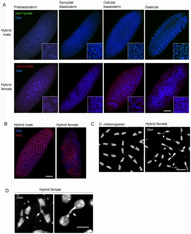
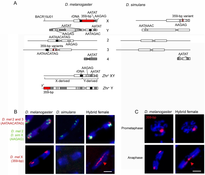
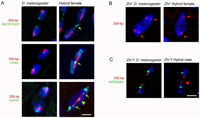
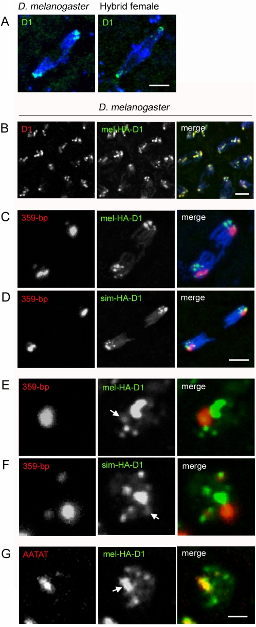
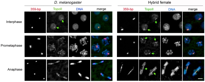
Postzygotic reproductive barriers such as sterility and lethality of hybrids are important for establishing and maintaining reproductive isolation between species. Identifying the causal loci and discerning how they interfere with the development of hybrids is essential for understanding how hybrid incompatibilities (HIs) evolve, but little is known about the mechanisms of how HI genes cause hybrid dysfunctions. A previously discovered Drosophila melanogaster locus called Zhr causes lethality in F1 daughters from crosses between Drosophila simulans females and D. melanogaster males. Zhr maps to a heterochromatic region of the D. melanogaster X that contains 359-bp satellite repeats, suggesting either that Zhr is a rare protein-coding gene embedded within heterochromatin, or is a locus consisting of the noncoding repetitive DNA that forms heterochromatin. The latter possibility raises the question of how heterochromatic DNA can induce lethality in hybrids. Here we show that hybrid females die because of widespread mitotic defects induced by lagging chromatin at the time during early embryogenesis when heterochromatin is first established. The lagging chromatin is confined solely to the paternally inherited D. melanogaster X chromatids, and consists predominantly of DNA from the 359-bp satellite block. We further found that a rearranged X chromosome carrying a deletion of the entire 359-bp satellite block segregated normally, while a translocation of the 359-bp satellite block to the Y chromosome resulted in defective Y segregation in males, strongly suggesting that the 359-bp satellite block specifically and directly inhibits chromatid separation. In hybrids produced from wild-type parents, the 359-bp satellite block was highly stretched and abnormally enriched with Topoisomerase II throughout mitosis. The 359-bp satellite block is not present in D. simulans, suggesting that lethality is caused by the absence or divergence of factors in the D. simulans maternal cytoplasm that are required for heterochromatin formation of this species-specific satellite block. These findings demonstrate how divergence of noncoding repetitive sequences between species can directly cause reproductive isolation by altering chromosome segregation.


Conflict of interest statement

The authors have declared that no competing interests exist.

Figures

Figure 1
Figure 1. Young hybrid female embryos exhibit defects in nuclear spacing, mitotic asynchrony, and lagging chromatin.
(A) Hybrid male and female embryos at different stages of early embryogenesis. Embryos were sexed using FISH probes to the D. melanogaster X (359-bp satellite) and Y (AATAGAC satellite) chromosomes. Higher magnifications of the nuclei are shown in the insets. Hybrid male embryos develop normally, while hybrid female embryos show abnormal nuclear spacing beginning during nuclear divisions 10–13 of the syncytial blastoderm stage. (B) Hybrid female embryos exhibit mitotic asynchrony. Left panel, a hybrid male embryo showing wild-type pattern. Right panel, a hybrid female embryo. Both embryos were stained with anti-phospho-Histone 3 (PH3) antibodies. (C) Chromosome mis-segregation in hybrid female embryos. White arrows in right panel indicate lagging chromatin during anaphase in a hybrid female embryo. (D) Left panel is a high magnification of two dividing chromosome sets during anaphase. Right panel is a high magnification of two daughter nuclei during late telophase. White arrows indicate lagging chromatin. Scale bar is 90 µm in (A) and (B), 8 µm in (C), and 5 µm in (D).
Figure 2
Figure 2. Separation failure of the D. melanogaster X chromatids during anaphase in hybrid female embryos.
(A) Schematic of satellites and other loci used as targets of FISH, and of Zhr mutant and duplicated chromosomes. Mapping of FISH probes is shown in Figure S2. Boxes and lines represent heterochromatin and euchromatin, respectively. Constrictions in boxes represent the centromeres. Chromosomal regions are not drawn to scale. The Zhr 1 chromosome is thought to have resulted from a recombination between an X and a Y . Our FISH analysis shows that this compound-XY chromosome contains the Y centromere and the proximal half of the Y long arm fused with the X euchromatin and distal pericentric heterochromatin (Figure S2); note that this structure differs from that inferred by Sawamura et al. . In this chromosome, the entire 359-bp satellite block has been deleted. The Zhr + Y chromosome was made by a translocation onto the distal end of the Y long arm of half of the X-derived 359-bp satellite block and a small amount of distal euchromatin carrying the y + marker from an inverted X chromosome (In(1)sc 8) . (B) Top row, D. melanogaster 2 and 3 chromatids and the D. simulans X chromatids segregate normally in hybrid female embryos. Bottom row, the D. melanogaster X chromatids, marked by a probe for the 359-bp satellite (red arrowhead), fail to segregate and comprise the lagging chromatin. (C) The 359-bp satellite block is normally condensed during prometaphase but abnormally stretched (red arrowhead) during anaphase in hybrid female embryos. DNA is blue in (B) and (C). Scale bar is 5 µm in (B) and (C).
Figure 3
Figure 3. Stretched and lagging X heterochromatin is induced by sequences in the 359-bp satellite block in hybrid female embryos.
(A) Green arrows in right top and middle panels indicate unstretched euchromatin and rDNA, respectively. In the right bottom panel, green arrows highlight AATAT satellite DNA proximal to the 359-bp DNA block that is segregating to the spindle poles, while the green arrowhead indicates a small amount of stretched and lagging AATAT DNA. (B) Normal X chromosome segregation in hybrid females carrying the Zhr 1 compound-XY chromosome, which is devoid of the X-linked 359-bp satellite. The related satellites (353-bp, 356-bp, and 361-bp repeats, indicated by red arrows) on Chromosome 3 segregate normally. (C) 359-bp DNA translocated to the Y chromosome is stretched and lagging in a hybrid male embryo (right panel). The D. melanogaster control embryo carries the Zhr 1 compound-XY chromosome instead of a wild-type X chromosome, so that the only source of 359-bp satellite is the Y. The red arrowhead points to lagging Y-linked 359-bp satellite DNA. DNA is blue in all panels. Scale bar is 5 µm in (A) and 7 µm in (C).
Figure 4
Figure 4. D1 localizes to AATAT satellite DNA but not the 359-bp satellite block during early embryogenesis.
(A) Left panel, an anaphase spindle in a D. melanogaster embryo. Anti-D1 highlights endogenous D1, which localizes to pericentromeric regions near the spindle poles. Right panel, an anaphase spindle in a hybrid female embryo. Endogenous D1 exhibits a pattern similar to that found in D. melanogaster. DNA is blue in both panels. (B) Anaphase spindles from a D. melanogaster embryo with HA-tagged D. melanogaster D1 (UAS-mel-HA-D1 driven by P{matα4-GAL-VP16}V37). Anti-D1 recognizes both endogenous and HA-D1. The two signals overlap completely, showing that the HA-D1 protein localizes identically to endogenous D1. (C) Anaphase spindle from the same genotype in (B), showing mel-HA-D1 and 359-bp DNA. (D) Anaphase spindle from a D. melanogaster embryo with HA-tagged D. simulans D1 (UAS-sim-HA-D1 driven by P{matα4-GAL-VP16}V37). Neither of the D1 orthologs, (C) and (D), co-localizes with the 359-bp satellite block. (E) Interphase nucleus from the same genotype in (C). (F) Interphase nucleus from the same genotype in (D). Both D1 orthologs show only a slight co-localization with the 359-bp satellite block during interphase. White arrows in (E) and (F) indicate this minimal co-localization. (G) Interphase nucleus from the same genotype in (C). The mel-HA-D1 protein co-localizes strongly with AATAT satellite DNA (indicated by white arrow). Scale bar is 4 µm in (A), 5 µm in (B), 4 µm in (C, D) and 3 µm in (E–G).
Figure 5
Figure 5. Topoisomerase II (TopoII) is mis-localized on the 359-bp satellite block during mitosis in hybrid female embryos.
Green arrows indicate TopoII localized to the 359-bp satellite block. Scale bar is 5 µm.

Comment in

References

    1. Ting C-T, Tsaur S-C, Wu M-L, Wu C-I. A rapidly evolving homeobox at the site of a hybrid sterility gene. Science. 1998;282:1501–1504. - PubMed
    1. Presgraves D. C, Balagopalan L, Abmayr S. M, Orr H. A. Adaptive evolution drives divergence of a hybrid inviability gene between two species of Drosophila. Nature. 2003;423:715–719. - PubMed
    1. Barbash D. A, Awadalla P, Tarone A. M. Functional divergence caused by ancient positive selection of a Drosophila hybrid incompatibility locus. PLoS Biol. 2004;2:e142. doi: 10.1371/journal.pbio.0020142. - DOI - PMC - PubMed
    1. Lee H-Y, Chou J-Y, Cheong L, Chang N-H, Yang S-Y, et al. Incompatibility of nuclear and mitochondrial genomes causes hybrid sterility between two yeast species. Cell. 2008;135:1065–1073. - PubMed
    1. Masly J. P, Jones C. D, Noor M. A, Locke J, Orr H. A. Gene transposition as a cause of hybrid sterility in Drosophila. Science. 2006;313:1448–1450. - PubMed

Publication types