Review
. 2009 Nov;9(5):507-17.
doi: 10.2174/187152609789105713.

Structural genomics and drug discovery for infectious diseases

Affiliations
Review

Structural genomics and drug discovery for infectious diseases

W F Anderson. Infect Disord Drug Targets. 2009 Nov.

Abstract

The application of structural genomics methods and approaches to proteins from organisms causing infectious diseases is making available the three dimensional structures of many proteins that are potential drug targets and laying the groundwork for structure aided drug discovery efforts. There are a number of structural genomics projects with a focus on pathogens that have been initiated worldwide. The Center for Structural Genomics of Infectious Diseases (CSGID) was recently established to apply state-of-the-art high throughput structural biology technologies to the characterization of proteins from the National Institute for Allergy and Infectious Diseases (NIAID) category A-C pathogens and organisms causing emerging, or re-emerging infectious diseases. The target selection process emphasizes potential biomedical benefits. Selected proteins include known drug targets and their homologs, essential enzymes, virulence factors and vaccine candidates. The Center also provides a structure determination service for the infectious disease scientific community. The ultimate goal is to generate a library of structures that are available to the scientific community and can serve as a starting point for further research and structure aided drug discovery for infectious diseases. To achieve this goal, the CSGID will determine protein crystal structures of 400 proteins and protein-ligand complexes using proven, rapid, highly integrated, and cost-effective methods for such determination, primarily by X-ray crystallography. High throughput crystallographic structure determination is greatly aided by frequent, convenient access to high-performance beamlines at third-generation synchrotron X-ray sources.


Figures
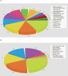
Fig. (1)
Fig. (1)
Current CSGID Organism Distribution: Targets and Structures. (A) Targets: Bacillus anthracis (544), Salmonella typhimurium (437), Yersinia pestis (406), Francisella tularensis (264), Vibrio cholerae (262), Staphylococcus aureus (261), Coxiella burnetii (166), Campylobacter jejuni (156), Listeria monocytogenes (93), Monkeypox virus (50), Cowpox virus (38), Shigella flexneri (27), Dengue virus (20), Escherichia coli (19), Myxoma virus (16), Clostridium difficile (7), others (18). (B) Structures: Salmonella typhimurium (19), Vibrio cholerae (14), Bacillus anthracis (11), Staphylococcus aureus (12), Yersinia pestis (11), Shigella flexneri (2), Dengue virus (1), Listeria monocytogenes (1), Francisella tularensis (1).
Fig. (2)
Fig. (2)
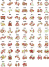
Structures of 67 unique targets solved by CSGID as May 2009. The PDB codes for these structures are: 3FZY [27], 3G1Z [38], 3FF1 [39], 3G25 [40], 3FTT [24], 3FWX [41], 3E9A [42], 3FWW [43], 3G48 [44], 3FPK [45], 3G0A [46], 3GOM [47], 3G0S [48], 3EEV [20], 3EFV [49], 3F0I [50], 3ERP [51], 3F09 [52], 3HL3 [95], 3EME [53], 3EFB [54], 3F6M [55], 3F4N [56], 3FOB [57], 3DQQ [58], 34EF [19], 3EDN [59], 3EFE [60], 3E7N [61], 3DR6 [21], 3H07 [62], 3EGJ [63], 3EC6 [64], 3EG7 [26], 3EGP [65], 3DZC [66], 3GRI [67], 3DR3 [68], 3ECT [22], 3ED6 [69], 3EER [70], 3GSD [71], 3GBX [72], 3GEU [73], 3GHZ [74], 3GJZ [75], 3GC2 [76], 3HJJ [25], 3GA7 [77], 3H1S [78], 3H0P [79], 3GO9 [80], 3H2Y [81], 3H5Q [82], 3H88 [83], 3H83 [84], 3GSE [85], 3H02 [86], 3CWC [87], 3HFR [88], 3GVD [23], 3HHO [89], 3HJB [90], 3GOS [91], 3HID [92], 3GIU [93], and 3HJV [96].
Fig. (3)
Fig. (3)
Structures from the menaquinone biosynthetic pathway determined by CSGID as of May 2009. Adapted from [94].
Fig. (4)
Fig. (4)
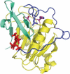
Structure of the cysteine protease domain (CPD) of the Vibrio cholerae MARTX. The CPD is activated after translocation into the host cell by the binding of inositol hexaphosphate (InsP6, red in the picture). CPD is a caspase-like protein that has the leucine (purple) of its N-terminal cleavage site bound in a hydrophobic specificity pocket and the scissile peptide bond located between the serine that was substituted for the essential cysteine (C3568S, catalytically inactive mutant) and the conserved histidine residue (both shown with magenta carbon atoms). The InsP6 activating ligand is bound by a number of positively charged lysine and arginine residues from the N-terminal region (blue), the main body of the CDP (yellow) and the C-terminal region (green). Related toxins with Cysteine Protease Domains, but other cytopathic domains, occur in a number of other gram-negative bacteria.

References

    1. Payne DJ, Gwynn MN, Holmes DJ, Pompliano DL. Nat. Rev. Drug Discov. 2007;6:29–40. - PubMed
    1. Taubes G. Science. 2008;321:356–361. - PubMed
    1. Alekshun MN, Levy SB. Cell. 2007;128:1037–1050. - PubMed
    1. Payne DJ. Science. 2008;321:1644–1645. - PubMed
    1. Barker JJ. Drug Discov. Today. 2006;11:391–404. - PubMed

Publication types