. 2009 Oct 27:10:130.
doi: 10.1186/1471-2202-10-130.

Progranulin is expressed within motor neurons and promotes neuronal cell survival

Affiliations

Progranulin is expressed within motor neurons and promotes neuronal cell survival

Cara L Ryan et al. BMC Neurosci. .

Abstract

Background: Progranulin is a secreted high molecular weight growth factor bearing seven and one half copies of the cysteine-rich granulin-epithelin motif. While inappropriate over-expression of the progranulin gene has been associated with many cancers, haploinsufficiency leads to atrophy of the frontotemporal lobes and development of a form of dementia (frontotemporal lobar degeneration with ubiquitin positive inclusions, FTLD-U) associated with the formation of ubiquitinated inclusions. Recent reports indicate that progranulin has neurotrophic effects, which, if confirmed would make progranulin the only neuroprotective growth factor that has been associated genetically with a neurological disease in humans. Preliminary studies indicated high progranulin gene expression in spinal cord motor neurons. However, it is uncertain what the role of Progranulin is in normal or diseased motor neuron function. We have investigated progranulin gene expression and subcellular localization in cultured mouse embryonic motor neurons and examined the effect of progranulin over-expression and knockdown in the NSC-34 immortalized motor neuron cell line upon proliferation and survival.

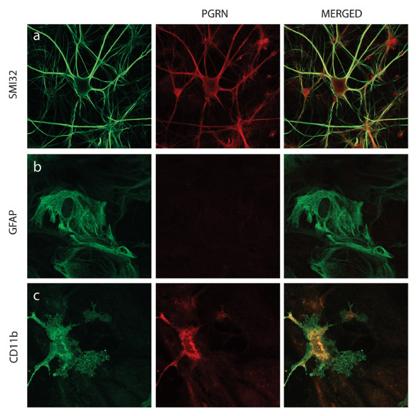
Results: In situ hybridisation and immunohistochemical techniques revealed that the progranulin gene is highly expressed by motor neurons within the mouse spinal cord and in primary cultures of dissociated mouse embryonic spinal cord-dorsal root ganglia. Confocal microscopy coupled to immunocytochemistry together with the use of a progranulin-green fluorescent protein fusion construct revealed progranulin to be located within compartments of the secretory pathway including the Golgi apparatus. Stable transfection of the human progranulin gene into the NSC-34 motor neuron cell line stimulates the appearance of dendritic structures and provides sufficient trophic stimulus to survive serum deprivation for long periods (up to two months). This is mediated at least in part through an anti-apoptotic mechanism. Control cells, while expressing basal levels of progranulin do not survive in serum free conditions. Knockdown of progranulin expression using shRNA technology further reduced cell survival.

Conclusion: Neurons are among the most long-lived cells in the body and are subject to low levels of toxic challenges throughout life. We have demonstrated that progranulin is abundantly expressed in motor neurons and is cytoprotective over prolonged periods when over-expressed in a neuronal cell line. This work highlights the importance of progranulin as neuroprotective growth factor and may represent a therapeutic target for neurodegenerative diseases including motor neuron disease.


Figures

Figure 1
Figure 1
PGRN is expressed by neurons of both central and peripheral nervous systems, in vivo and in vitro in the mouse. Gene expression pattern of murine PGRN in brain (B, C); cervical spinal cord (A, D, E) and primary cultures of dissociated spinal cord-DRG (F, G). In situ hybridization, to detect PGRN mRNA in saggital section of pontine grey matter (A) and cross-section of cervical spinal cord (D, E). The majority of neurons throughout the grey matter of the spinal cord express PGRN as well as ependymal cells and possibly microglial cells (A). Note in particular the robust expression of PGRN mRNA in large motor neurons in panels B, D. Panels C and E illustrate the hybridization signal observed with the sense control applied to serial sections to those shown in panels B and D, respectively. (F) Motor neurons (asterisk) as well as other neuronal subtypes in dissociated spinal cord-DRG cultures, express PGRN; (G) equivalent sense control. Scale bar (panels B-G) represents 20 μm. Original magnification of panel A was 10×
Figure 2
Figure 2
PGRN is localized within motor neurons of the mouse lumbar spinal cord. PGRN is localized within motor neurons of the mouse spinal cord. Labelling of paraffin-fixed cross-sections of murine spinal cord with SMI32 marker against hypo-phosphorylated neurofilaments (left panel), which is a marker for motor neurons, and anti-PGRN (middle panel). Merged channels are shown in the right panel. (a) at original magnification (40×), (b) at magnification (63×).
Figure 3
Figure 3
PGRN expression within the dissociated spinal cord-DRG cultures. Confocal Images taken of dissociated spinal cord cultures. PGRN (red) is very clearly expressed within motor neurons (a), labelled with SMI32 (green). PGRN is also expressed by microglia, as demonstrated by colocalization between PGRN and CD11b (c). Astrocytes, however, do not express PGRN, as demonstrated in (b).
Figure 4
Figure 4
PGRN within motor neurons in primary cultures does not colocalize with the nucleus or mitochondria. (A) Motor neuron labeled with antibody to mouse PGRN (left hand image) is attenuated by antigen-competition with 300 ng recombinant mouse PGRN (middle and right hand images). When anti-PRGN was pre-absorbed with 400 ng of mouse recombinant PGRN, no signal was observed in the primary motor neurons (not shown). Shown are confocal images taken at 100×. (B) PGRN is not distributed in nuclei or mitochondria, organelles that are not part of the secretory pathway. Immunolabelling of motor neurons in dissociated spinal cord-DRG cultures with anti-TDP-43 (a) and anti-cytochrome C (b) and anti-PGRN (middle column). Merged images (right column) show no colocalization of TDP-43 or cytochrome C with endogenous mouse PGRN. Confocal images were captured at 63× magnification, hatched boxes represent 3-5× zoom.
Figure 5
Figure 5
Subcellular localization of PGRN within motor neurons in primary cultures relative to markers for the ER, Golgi apparatus and chromogranin-A containing vesicles. Immunolabelling of motor neurons in dissociated spinal cord-DRG cultures with anti-Calreticulin (a), anti-GM130 (b), Chromogranin A (c) and anti-PGRN (middle column). Confocal images were captured at 63× magnification, hatched boxes represent 3-5× zoom.
Figure 6
Figure 6
Subcellular localization of PGRN within motor neurons in primary cultures relative to a marker for the lysosomal compartment markers for neurotransmitter vesicle trafficking and release (SNAP-25 and synaptophysin). Immunolabelling of motor neurons in dissociated spinal cord-DRG cultures with LysoTracker™ (a), SNAP-25 (b), Synaptophysin (c), and anti-PGRN (middle column). Confocal images were captured at 63× magnification, hatched boxes represent 3-5× zoom.
Figure 7
Figure 7
pEGFP-N1-hPGRN cloning construct. The human PGRN cDNA (hpgrn) was cloned into the pEGFP-N1 plasmid using EcoR1 and Sal1 restriction enzyme digestion sites. The eGFP molecule is fused to the C-terminus of the PGRN protein, and therefore does not affect its N-terminal signal sequence that is required for entry into the endoplasmic reticulum. Individual 12 cysteine granulin modules are designated 1 through to 7 and A through to G. P is paragranulin, a half-granulin module bearing six cysteine residues.
Figure 8
Figure 8
Transfection of pEGFP-N1-hPGRN plasmid shows granular morphology and particular PGRN localization in Golgi. The overlap seen with PGRN and the Golgi marker GM130 (a) is very prominent, while there is no overlap between mitochondria (b) and PGRN. Panel (c) reveals the pEGFP-N1 vector only transfection, having primarily nuclear GFP signal, and a very different subcellular distribution of PGRN, which appears distinctly granular in panels (a and b). Confocal images were taken at 63×.
Figure 9
Figure 9
Validation of Stable Transfectants. The motor neuron-neuroblastoma hybrid cell line, NSC-34, was transfected with empty pcDNA vector or pcDNA-hPGRN and selected for drug resistance over a three-week period. (A) Species-specific PGRN primers were used to confirm the stable integration of hPGRN. (B) Western Blot analysis confirmed presence of hPGRN in stable transfectants and not in the vector only control cells.
Figure 10
Figure 10
PGRN over-expression in NSC-34 cells promotes a neuronal morphology. Untransfected NSC-34 cells (A-D), transfected with pcDNA3 vector only (E-H) or transfected with pcDNA3-hPGRN (I-L). Micrographs showing the distribution of DAPI (A, E, I), F-actin (B, F, J), hPGRN (C, G, K) and merged images (D, H, L). PGRN over-expression promotes more extensive cytoskeletal extensions (hatched box) and is localized within presumptive secretory granules (arrowheads).
Figure 11
Figure 11
PGRN is a sufficient trophic stimulus to maintain prolonged survival of NSC-34 cells in serum-free medium. NSC-34 cells stably transfected with pcDNA3/PGRN and grown in serum-free medium (A) for 20 days in six well plates, the cut-out box illustrating the same cells photographed 3 hr later showing continued active extension and retraction of processes; (B) for 51 days, and (C, D) for 67 days in serum free medium. All NSC-34/vector control cells died before day 20 (not shown). Images were taken at an original magnification of 15×.
Figure 12
Figure 12
PGRN over-expression prevents apoptosis of NSC-34 cells induced by serum deprivation and exogenous PGRN increases cell survival. Stable vector only transfectants (pcDNA, open bars) and cells stably over-expressing hPGRNs (pcDNA-hPGRN; black bars) were cultured in serum-free RPMI medium in six well plates. (A) Number of cells per well were determined at three-day intervals for fifteen days. NSC-34 cells that over-expressed hPGRN demonstrated increased survival as compared to controls (N = 2 6 fields 10× magnification/each condition, Asterisks denote P < 0.005). (B) Cell proliferation assay based on 18 hr BrdU incorporation following 1, 3 and 5 days culture in serum-free medium in 96 well plates. Over-expression of hPGRN during serum deprivation did not significantly increase cell proliferation rates (10 replicates per measurement P > 0.1). (C) Apoptosis assay based on the TUNEL- labelling method following 6 days in serum-free medium. Over-expression of hPGRN during serum deprivation protected against apoptosis (N = 2 6 fields 10× magnification per condition. Asterisks denote P < 0.0001). (D) Addition of exogenous PGRN also dramatically increased NSC-34 survival (P < 0.0001).
Figure 13
Figure 13
Short hairpin RNA silencing of PGRN in NSC 34. (A) RT-PCR of PGRN and actin in NSC34 cells stably -transfected with shRNA. (B) Relative expression ratio of quantitative RT-PCR showing the expression of PGRN mRNA in NSC34/pRS plasmid and NSC34/shPGRN. (C) Western blot analysis of PGRN and actin in NSC34 cells with stably-transfected with shRNA.
Figure 14
Figure 14
PGRN knockdown reduces cell proliferation and exogenous PGRN rescues cell proliferation. (A) AlamarBlue cell proliferation assay allowing the quantitative measurement of cell proliferation from day 0 to day7 in the presence of serum in 96 well plates. PGRN knockdown significantly reduced the cell proliferation rate from day 2-3 to day 7-8 (10 replicates per determination, p > 0.001). (B) Addition of exogenous PGRN rescued NSC-34 cell proliferation from day 2-3 to 4-5 (representative experiment, 10 replicated per determination, P < 0.05). (C) Apoptosis assay based on TUNEL labelling method following 3 days in the presence of serum. PGRN silencing had no significant effect on apoptosis (N = 2, 6 fields 10× magnification per determination).

References

    1. Bhandari V, Palfree RG, Bateman A. Isolation and sequence of the granulin precursor cDNA from human bone marrow reveals tandem cysteine-rich granulin domains. Proc Natl Acad Sci USA. 1992;89(5):1715–1719. doi: 10.1073/pnas.89.5.1715. - DOI - PMC - PubMed
    1. Plowman GD, Green JM, Neubauer MG, Buckley SD, McDonald VL, Todaro GJ, Shoyab M. The epithelin precursor encodes two proteins with opposing activities on epithelial cell growth. The Journal of Biological Chemistry. 1992;267:13073–13078. - PubMed
    1. Zhou J, Gao G, Crabb JW, Serrero G. Purification of an autocrine growth factor homologous with mouse epithelin presursor from a highly tumorigenic cell line. The Journal of Biological Chemistry. 1993;268:10863–10869. - PubMed
    1. Anakwe OO, Gerton GL. Acrosome biogenesis begins during meiosis: evidence from the synthesis and distribution of an acrosomal glycoprotein, acrogranin, during guinea pig spermatogenesis. Biology of Reproduction. 1990;42:317–328. doi: 10.1095/biolreprod42.2.317. - DOI - PubMed
    1. Ong CH, Bateman A. Progranulin (granulin-epithelin precursor, PC-cell derived growth factor, acrogranin) in proliferation and tumorigenesis. Histology and Histopathology. 2003;18:1275–1288. - PubMed

Publication types

MeSH terms

LinkOut - more resources