. 2009 Dec;15(12):2312-20.
doi: 10.1261/rna.1584209. Epub 2009 Oct 27.

Structural features of the tmRNA-ribosome interaction

Affiliations

Structural features of the tmRNA-ribosome interaction

Elizaveta Y Bugaeva et al. RNA. 2009 Dec.

Abstract

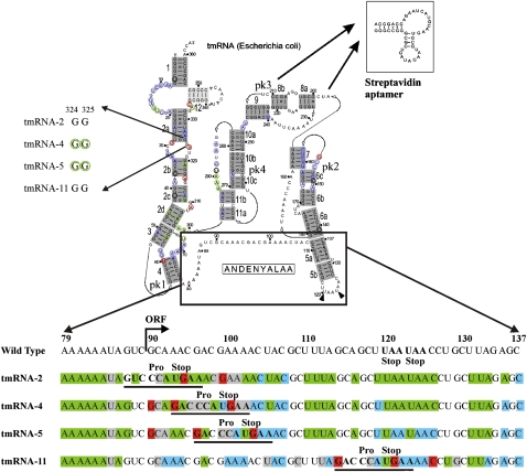
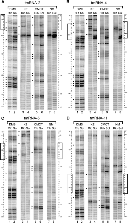
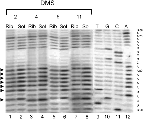
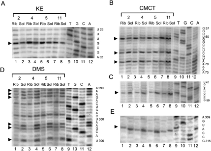
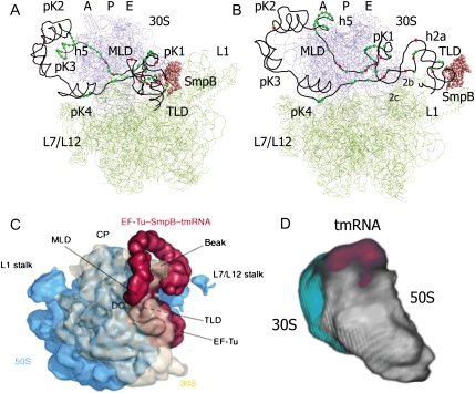
Trans-translation is a process which switches the synthesis of a polypeptide chain encoded by a nonstop messenger RNA to the mRNA-like domain of a transfer-messenger RNA (tmRNA). It is used in bacterial cells for rescuing the ribosomes arrested during translation of damaged mRNA and directing this mRNA and the product polypeptide for degradation. The molecular basis of this process is not well understood. Earlier, we developed an approach that allowed isolation of tmRNA-ribosomal complexes arrested at a desired step of tmRNA passage through the ribosome. We have here exploited it to examine the tmRNA structure using chemical probing and cryo-electron microscopy tomography. Computer modeling has been used to develop a model for spatial organization of the tmRNA inside the ribosome at different stages of trans-translation.


Figures

FIGURE 1.
FIGURE 1.
Protection pattern of tmRNA in ribosomal complexes. The secondary structure of tmRNA was adapted from data from rnp.uthct.edu/rnp/tmRDB/tmRDB.html. pk3 (nucleotides U212–A239) was substituted with an aptamer to streptavidin (inset). The effects that are similar for all complexes are shown at the secondary structure as a green circle for the protection, red for the exposure, blue for the nucleotides which are equally exposed for the modification both in the solution and in the complexes. Black circle indicates nucleotides that are inclined for degradation. The nucleotides 324GG325 which displayed different accessibility to the modifying reagents in different complexes are shown at the left side. The region A79–C137 is shown in details at the bottom. Protected nucleotides are colored in green, exposed in red, nucleotides which are equally exposed for the modification both in the solution and in the complexes are in blue, and nucleotides inclined for degradation are in gray.
FIGURE 2.
FIGURE 2.
Chemical probing of the MLD and helix 5 tmRNA region in the ribosomal complexes (Rib) and in the solution (Sol) for tmRNA-2 (A), tmRNA-4 (B), tmRNA-5 (C), and tmRNA-11 (D) by DMS, KE, and CMCT. NM, nonmodified controls. The sequence of tmRNA is shown on the sides of the gels. The mutated sequences of the stop signal are boxed. Arrows indicate the position of nucleotides which displayed different accessibility to modifying reagents in the complexes and in solution.
FIGURE 3.
FIGURE 3.
Chemical probing of the A79–A86 tmRNA region in the ribosomal complexes (Rib) and in the solution (Sol) for tmRNA-2 (2), tmRNA-4 (4), tmRNA-5 (5), and tmRNA-11 (11) by DMS. Dideoxy sequencing lanes are indicated by T, G, C, and A. The sequence of tmRNA is shown on the right side of the gels. Arrows indicate the position of nucleotides which displayed different accessibility to modifying reagents in the complexes and in solution.
FIGURE 4.
FIGURE 4.
Chemical probing of the pK1, pK4, and helix 2 tmRNA region in the ribosomal complexes (Rib) and in the solution (Sol) for tmRNA-2 (2), tmRNA-4 (4), tmRNA-5 (5), and tmRNA-11 (11) by KE (A), CMCT (B,C,E), and DMS (D). Dideoxy sequencing lanes are indicated by T, G, C, and A. The sequence of tmRNA is shown on the right side of the gels. Arrows indicate the position of nucleotides which displayed different accessibility to modifying reagents in the complexes and in solution.
FIGURE 5.
FIGURE 5.
Chemical probing of the helix 2 (A) and pK2 (B) of tmRNA in the ribosomal complexes (Rib) and in the solution (Sol) for tmRNA-2 (2), tmRNA-4 (4), tmRNA-5 (5), and tmRNA-11 (11) by DMS and KE. Dideoxy sequencing lanes are indicated by T, G, C, and A. The sequence of tmRNA is shown on the right side of the gels. Arrows indicate the position of nucleotides which displayed different accessibility to modifying reagents in the complexes and in solution.
FIGURE 6.
FIGURE 6.
Chemical probing of the TLD of tmRNA in the ribosomal complexes (Rib) and in the solution (Sol) for tmRNA-2 (2), tmRNA-4 (4), tmRNA-5 (5), and tmRNA-11 (11) by CMCT (A) and DMS (B). Dideoxy sequencing lanes are indicated by T, G, C, and A. The sequence of tmRNA is shown on the right side of the gels. Arrows indicate the position of nucleotides which displayed the different accessibility to modifying reagents in the complexes and in solution.
FIGURE 7.
FIGURE 7.
Structural models of tmRNA-2 (A) and tmRNA-4 (B) in complex with the ribosome (top views). 30S ribosomal subunit is colored in blue, 50S in green. tmRNA is shown as a black line connecting the phosphorus atoms. SmpB is presented as rose spheres of Cα atoms. Nucleotides of tmRNA protected by the ribosome from the chemical modification are shown as green spheres. Nucleotides that became more accessible to modification after the complex formation are shown as red spheres. Structural elements of tmRNA are marked. (C) Cryo-EM structure of the pre-initiation tmRNA–ribosomal complex adapted with permission (from Elsevier © 2004, Haebel et al. 2004). SmpB–tmRNA–EF-Tu (red) emerging from the intersubunit space between the 50S subunit (blue) and 30S subunit (cream). (D) Cryo-electron tomography structure of elongating tmRNA–ribosomal complex. 30S ribosomal subunit is colored in light gray, 50S in light blue, and tmRNA in magenta.

References

    1. Adrian M, Dubochet J, Lepault J, McDowall AW. Cryo-electron microscopy of viruses. Nature. 1984;308:32–36. - PubMed
    1. Barends S, Wower J, Kraal B. Kinetic parameters for tmRNA binding to alanyl-tRNA synthetase and elongation factor Tu from Escherichia coli. Biochemistry. 2000;39:2652–2658. - PubMed
    1. Barends S, Karzai AW, Sauer RT, Wower J, Kraal B. Simultaneous and functional binding of SmpB and EF-Tu•GTP to the alanyl acceptor arm of tmRNA. J Mol Biol. 2001;314:9–21. - PubMed
    1. Bessho Y, Shibata R, Sekine S, Murayama K, Higashijima K, Hori-Takemoto C, Shirouzu M, Kuramitsu S, Yokoyama S. Structural basis for functional mimicry of long-variable-arm tRNA by transfer-messenger RNA. Proc Natl Acad Sci. 2007;104:8293–8298. - PMC - PubMed
    1. Bjornsson A, Isaksson LA. Accumulation of a mRNA intermediate by ribosomal pausing at a stop codon. Nucleic Acids Res. 1996;24:1753–1757. - PMC - PubMed

Publication types

MeSH terms

LinkOut - more resources