. 2009 Nov 15;15(22):6912-20.
doi: 10.1158/1078-0432.CCR-09-1698. Epub 2009 Oct 27.

Differential cellular responses to prolonged LDR-IR in MLH1-proficient and MLH1-deficient colorectal cancer HCT116 cells

Affiliations

Differential cellular responses to prolonged LDR-IR in MLH1-proficient and MLH1-deficient colorectal cancer HCT116 cells

Tao Yan et al. Clin Cancer Res. .

Abstract

Purpose: MLH1 is a key DNA mismatch repair (MMR) protein involved in maintaining genomic stability by participating in the repair of endogenous and exogenous mispairs in the daughter strands during S phase. Exogenous mispairs can result following treatment with several classes of chemotherapeutic drugs, as well as with ionizing radiation. In this study, we investigated the role of the MLH1 protein in determining the cellular and molecular responses to prolonged low-dose rate ionizing radiation (LDR-IR), which is similar to the clinical use of cancer brachytherapy.

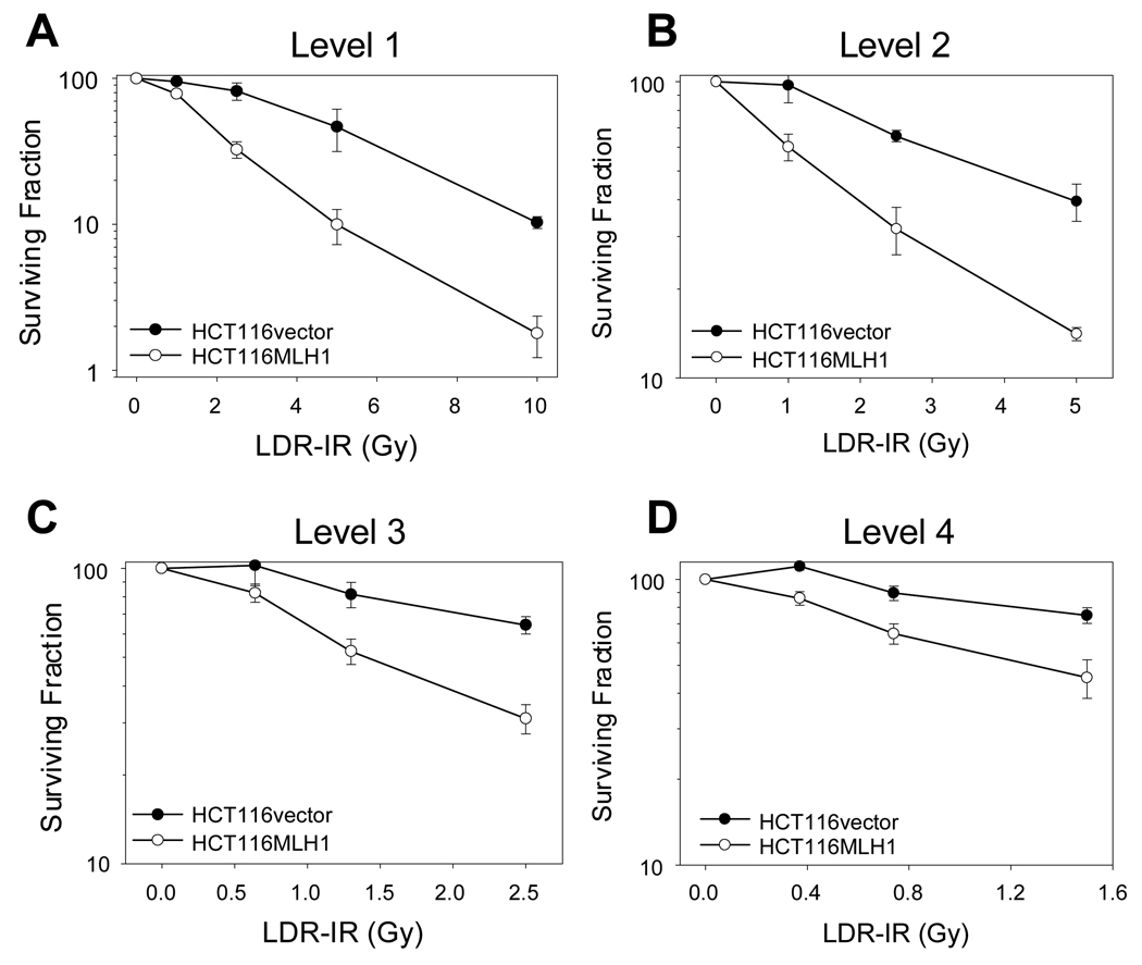
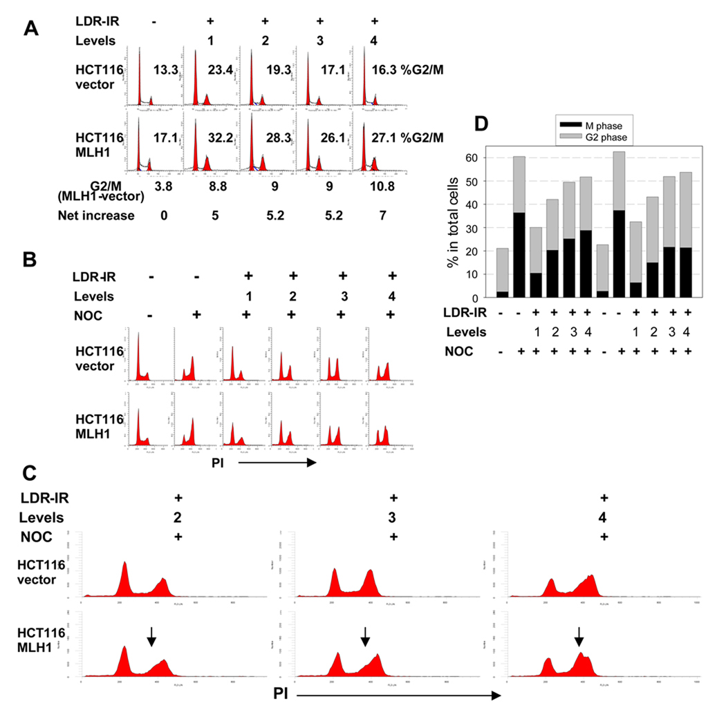
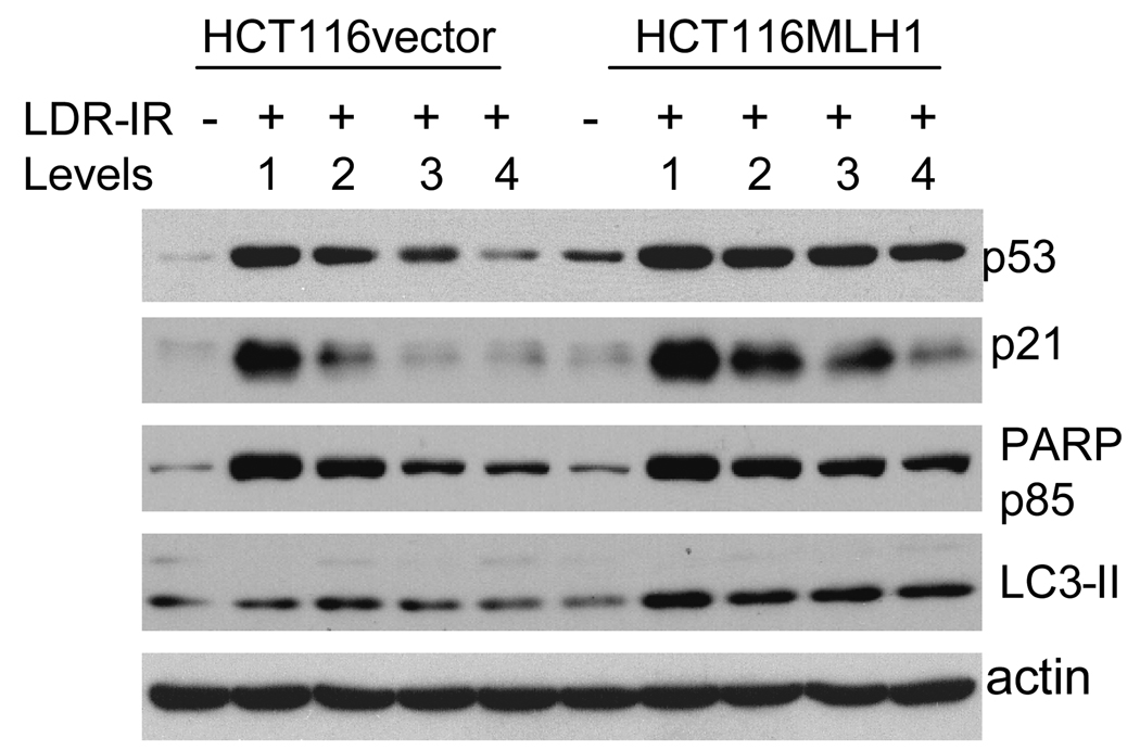
Experimental design: An isogenic pair of MMR(+) (MLH1(+)) and MMR(-) (MLH1(-)) human colorectal cancer HCT116 cells was exposed to prolonged LDR-IR (1.3-17 cGy/h x 24-96 h). The clonogenic survival and gene mutation rates were examined. Cell cycle distribution was analyzed with flow cytometry. Changes in selected DNA damage repair proteins, DNA damage response proteins, and cell death marker proteins were examined with Western blotting.

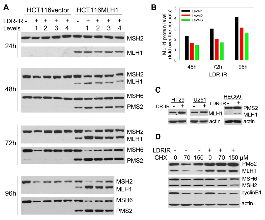
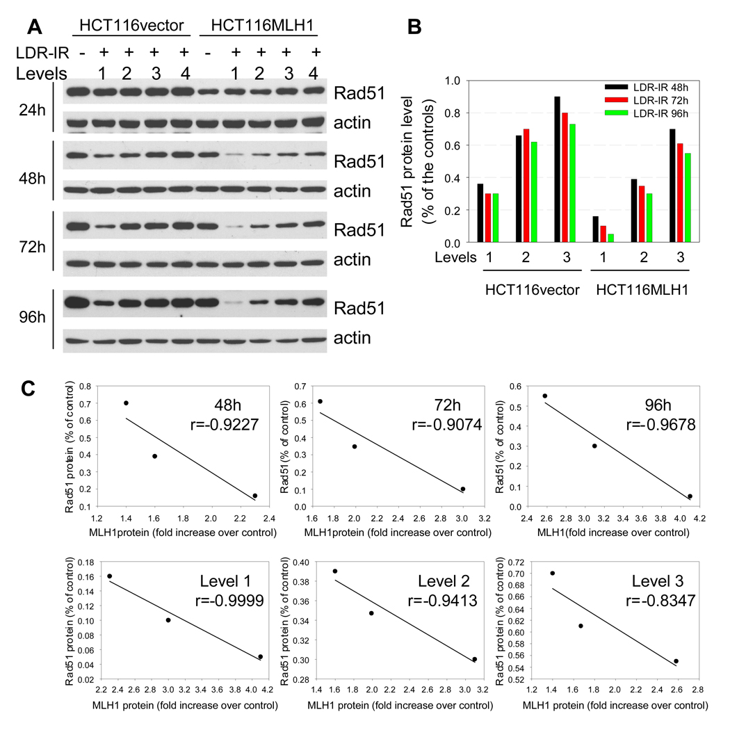
Results: MLH1(+) HCT116 cells showed greater radiosensitivity with enhanced expression of apoptotic and autophagic markers, a reduced HPRT gene mutation rate, and more pronounced cell cycle alterations (increased late-S population and a G(2)/M arrest) following LDR-IR compared with MLH1(-) HCT116 cells. Importantly, a progressive increase in MLH1 protein levels was found in MLH1(+) cells during prolonged LDR-IR, which was temporally correlated with a progressive decrease in Rad51 protein (involved in homologous recombination) levels.

Conclusions: MLH1 status significantly affects cellular responses to prolonged LDR-IR. MLH1 may enhance cell radiosensitivity to prolonged LDR-IR through inhibition of homologous recombination (through inhibition of Rad51).


Conflict of interest statement

Disclosure of Potential Conflicts of Interest: No potential conflicts of interest are disclosed.

Figures

Figure 1
Figure 1
MLH1 status affects cell sensitivity to prolonged LDR-IR. HCT116vector and HCT116MLH1 cells were exposed to prolonged LDR-IR simultaneously. A. Level 1, 9.3–11.1cGy/h. B, Level 2, 4.2–5.2 cGy/h. C, Level 3, 2.4–2.9 cGy/h. D, Level 4, 1.3–1.7 cGy/h. The data are from three experiments with significance at the P < 0.05 level.
Figure 2
Figure 2
HCT116MLH1 (MLH1+) cells demonstrate a greater late S population and a greater G2/M checkpoint arrest during prolonged LDR-IR than HCT116vector (MLH1) cells. A. flow cytometry histograms show moderately increased G2/M population after 72 h LDR-IR in both cell lines with about 5 percentage points more G2/M fraction in the MLH1+ cells than in the MLH1 cells. B. flow cytometry histograms show inhibition of G1/S progression in both cell lines. The cells were exposed to LDR-IR for 72 h and NOC was added for the last 8 h of LDR-IR prior to harvesting. C. the expanded portion of Fig. 2B demonstrates a late S phase population (arrows) in the MLH1+ cells. D. Bar graph derived from the representative scatter plots of dual-parameter flow cytometry for phospho-histone H3 (Ser10) and PI showing that the MLH1+ cells have less mitotic cells than the MLH1 cells. All experiments were repeated at least twice.
Figure 3
Figure 3
MLH1 protein accumulates in MLH1+ cells during prolonged LDR-IR as a result of compromised protein degradation. A. Western blots show that the increase in MLH1 and PMS2 proteins occurs concomitantly with decrease in MSH2/MSH6 proteins in HCT116MLH1 cells. B. MLH1 protein levels in Fig. 3A were quantified with Image J. C. Increased MLH1 protein can be seen in other MLH1+ cell lines HT29, U251 and HEC59 after LDR-IR (3.1 cGy/h for 72h). D. Protein synthesis inhibitor CHX cannot change the LDR-IR induced MLH1 accumulation (2.4 cGy/h for 96 h) in HCT116MLH1 cells. Cyclin B1 serves as a positive control, whereas PMS2, MSH2 and MSH6 are the negative controls. All Western blotting assays were repeated at least twice.
Figure 4
Figure 4
Rad51 protein decreases during prolonged LDR-IR. A. Western blots demonstrate that Rad51 protein decreases progressively during prolonged LDR-IR with more significant reduction found in HCT116MLH1 (MLH1+) cells. B. Rad51 protein levels in Fig. 4A were quantified with software Image J. C. Linear regression test shows a strong correlation between MLH1 protein increase and Rad51 protein decrease in the MLH1+ cells. Correlation coefficients are calculated on data sets from Fig. 4B using Excel statistics. All Western blotting assays are repeated at least twice.
Figure 5
Figure 5
Western blots demonstrate that levels of p53 and p21 proteins as well as cleaved PARP p85 fragment, are all elevated in both cell lines after a 72h exposure to prolonged LDR-IR but to a greater extent in the MLH1+ cells, while LC3-II is increased in the MLH1+ cells but not in the MLH1 cells. All Western blotting assays are repeated at least twice.

References

    1. Cardis E, Vrijheid M, Blettner M, et al. The 15-Country Collaborative Study of Cancer Risk among Radiation Workers in the Nuclear Industry: estimates of radiation-related cancer risks. Radiat Res. 2007;167:396–416. - PubMed
    1. Karagiannis TC, El-Osta A. Double-strand breaks: signaling pathways and repair mechanisms. Cell Mol Life Sci. 2004;61:2137–2147. - PMC - PubMed
    1. Georgakilas AG. Processing of DNA damage clusters in human cells: current status of knowledge. Mol Biosyst. 2008;4:30–35. - PubMed
    1. Yan T, Schupp JE, Hwang HS, et al. Loss of DNA mismatch repair imparts defective cdc2 signaling and G(2) arrest responses without altering survival after ionizing radiation. Cancer Res. 2001;61:8290–8297. - PubMed
    1. Kuraoka I, Bender C, Romieu A, Cadet J, Wood RD, Lindahl T. Removal of oxygen free-radical-induced 5',8-purine cyclodeoxynucleosides from DNA by the nucleotide excision-repair pathway in human cells. Proc Natl Acad Sci U S A. 2000;97:3832–3837. - PMC - PubMed

Publication types