. 2009 Nov 15;15(22):6830-40.
doi: 10.1158/1078-0432.CCR-09-0371. Epub 2009 Oct 27.

Tumor-derived tissue factor-bearing microparticles are associated with venous thromboembolic events in malignancy

Affiliations

Tumor-derived tissue factor-bearing microparticles are associated with venous thromboembolic events in malignancy

Jeffrey I Zwicker et al. Clin Cancer Res. .

Abstract

Purpose: Despite the strong association between malignant disease and thromboembolic disorders, the molecular and cellular basis of this relationship remains uncertain. We evaluated the hypothesis that tumor-derived tissue factor-bearing microparticles in plasma contribute to cancer-associated thrombosis.

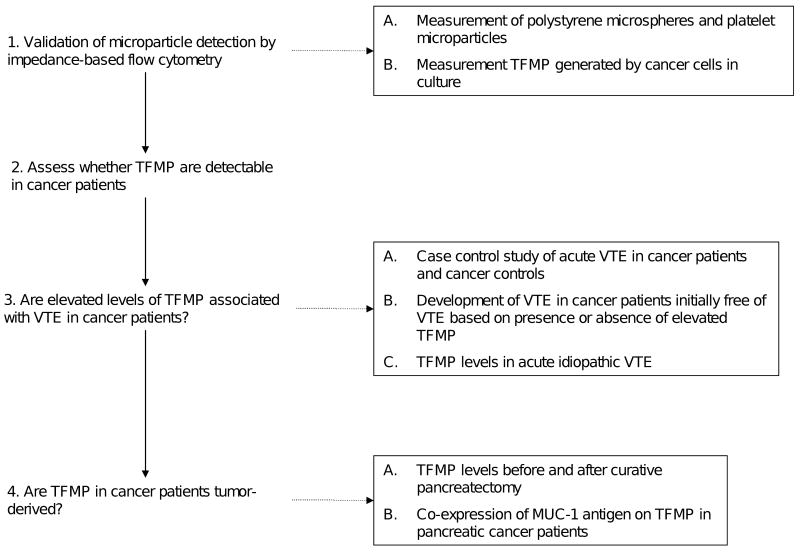
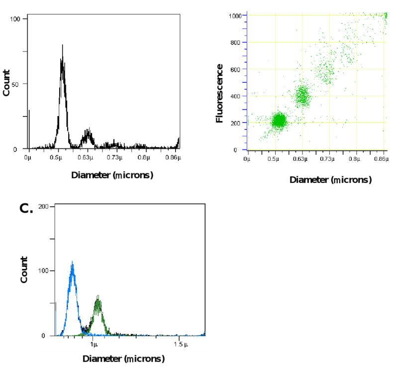
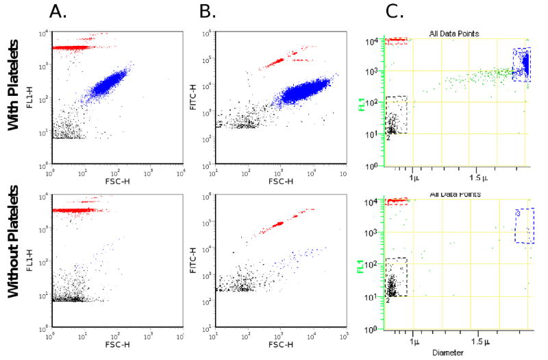
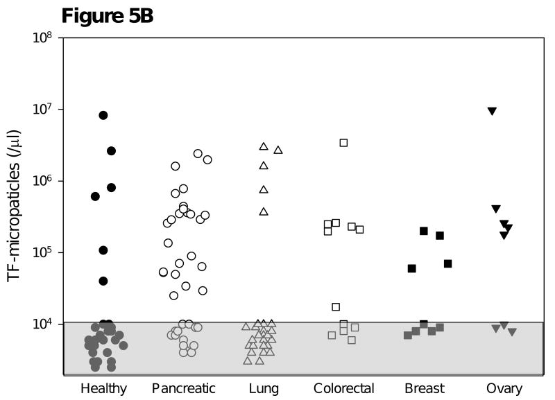
Experimental design: We developed impedance-based flow cytometry to detect, quantitate, and size microparticles in platelet-poor plasma. We evaluated the number of tissue factor-bearing microparticles in a cohort of cancer patients of different histologies (N = 96) and conducted a case-control study of 30 cancer patients diagnosed with an acute venous thromboembolic event (VTE) compared with 60 cancer patients of similar age, stage, sex, and diagnosis without known VTE, as well as 22 patients with an idiopathic VTE.

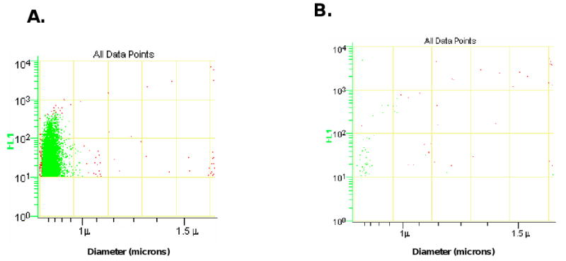
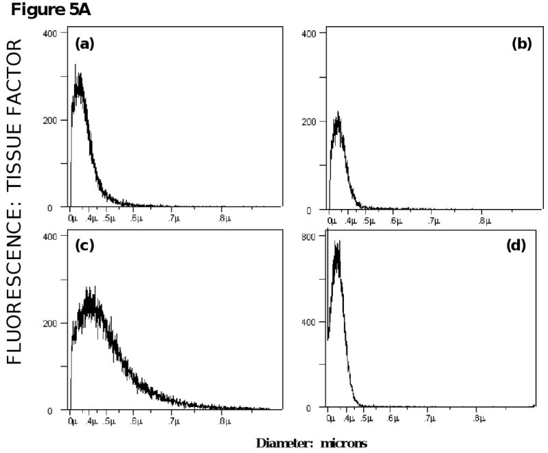
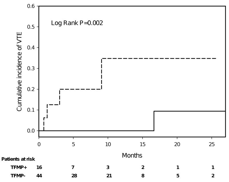
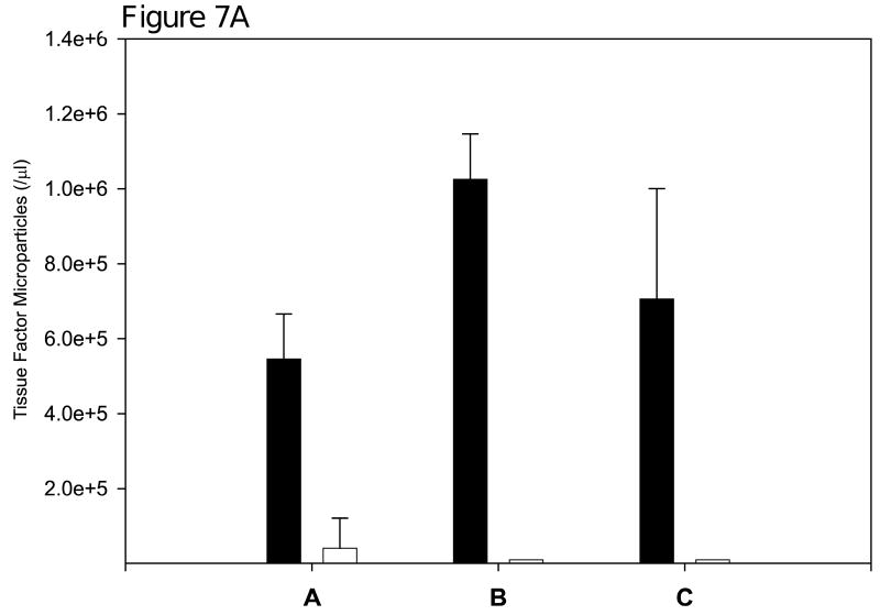
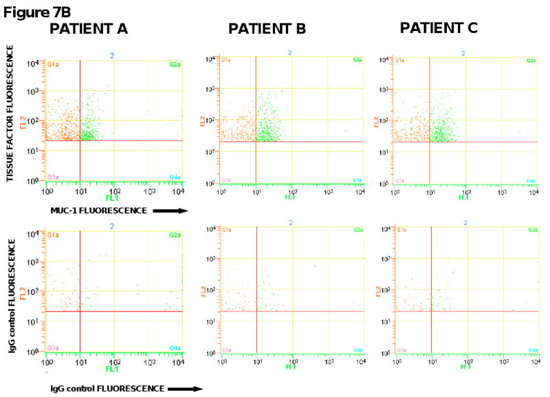
Results: Tissue factor-bearing microparticles were detected in patients with advanced malignancy, including two thirds of patients with pancreatic carcinoma. Elevated levels of tissue factor-bearing microparticles were associated VTE in cancer patients (adjusted odds ratio, 3.72; 95% confidence interval, 1.18-11.76; P = 0.01). In cancer patients without VTE, a retrospective analysis revealed a 1-year cumulative incidence of VTE of 34.8% in patients with tissue factor-bearing microparticles versus 0% in those without detectable tissue factor-bearing microparticles (Gray test P = 0.002).The median number of tissue factor-bearing microparticles in the cancer VTE cohort (7.1 x 10(4) microparticles/microL) was significantly greater than both the idiopathic VTE and cancer-no VTE groups (P = 0.002 and P = 0.03, respectively). Pancreatectomy in three patients eliminated or nearly eliminated these microparticles which coexpressed the epithelial tumor antigen, MUC-1.

Conclusion: We conclude that tumor-derived tissue factor-bearing microparticles are associated with VTE in cancer patients and may be central to the pathogenesis of cancer-associated thrombosis.


Figures

Figure 1
Figure 1
Flow diagram of studies performed to validate microparticle detection by impedance based flow cytometry and investigate the association between tumor-derived tissue factor bearing microparticles and VTE in cancer patients. (TFMP – tissue factor bearing microparticles)
Figure 2
Figure 2
Analysis of fluorescent microspheres using impedance-based flow cytometry. A. Histogram of 0.52 μm polystyrene fluorescent microspheres showing particle distribution as a function of particle size. B. Dot plot of particle diameter versus relative fluorescence for 0.52 μm microspheres. Some aggregates were observed. C. Histograms generated by measurement of electronic volumes of a mixed population of microspheres with diameters of 0.78 μm (blue) and 1.01 μm (green) microspheres showing full resolution of the two populations. Fluorescence was monitored at 488 nm.
Figure 3
Figure 3
Particle size comparisons. Platelet microparticles, platelets and 0.78 μm fluorescent microspheres were analyzed using impedance-based and light scatter-based flow cytometry (top panel). Platelets were removed by centrifugation; platelet microparticles and 0.78 μm microspheres alone were detected (bottom panel). A. Light scatter flow cytometry with Becton Dickinson FACSCalibur. Platelets (blue) and platelet microparticles (black) were partially resolved and both overlap with 0.78 μm microspheres (red). The platelet microparticles appear within the tail of the platelet population (x-axis forward scatter, y-axis fluorescence). B. Light scatter flow cytometry with the Becton Dickinson LSRII. Platelets (blue) and platelet microparticles (black) were partially resolved, and both overlap with the population of 0.78 μm microspheres (red) (x-axis forward scatter, y-axis fluorescence). C. Impedance-based flow cytometry. Platelets (blue) and platelet microparticles (black) were resolved (x-axis, diameter; y-axis, fluorescence), and can be compared to the 0.78 μm microspheres (red). Platelets and platelet microparticles were labeled with CD41a-FITC antibody. Platelets (blue), platelet microparticles (blue), and platelet microparticles (black). Histograms depict events accumulated over 30 seconds.
Figure 4
Figure 4
Tissue factor-bearing microparticles were measured in the conditioned media from the ASPC-1 pancreatic cancer cell line. The x-axis presents particle diameter and the y-axis presents fluorescence using a humanized anti-TF monoclonal antibody. The scatter plots for anti-tissue factor-Alexa 488 (A) and IgG-Alexa 488 control (B) are shown.
Figure 5
Figure 5
Survey of tissue factor-bearing microparticles in cancer patients. A. Histograms of tissue factor-bearing microparticle detected in four cancer patients. Tissue factor microparticle count versus particle diameter; the X axis is linear for particle volume but is presented in particle diameter in micrometers (0 to 1.0 μm). The patients had advanced disease and samples were drawn prior to a cycle of chemotherapy. The cancer diagnoses were (a) breast (b) pancreatic (c) pancreatic and (d) non-small cell lung cancer. Microparticle size varied from 0.3 μm (the lower limits of detection) to 500-800 μm. B. Tissue factor-bearing microparticle concentrations in advanced malignant disease. The percentage of patients with elevated levels of tissue factor bearing microparticles was significantly greater for pancreatic carcinoma (25 of 39) and colorectal carcinoma (7 of 12) compared with cancer-free subjects (6 of 31, P<.001 and P=0.03 respectively). Significant differences were not observed between cancer-free subjects and non-small cell lung carcinoma (5 of 28), breast carcinoma (4 of 9) and ovarian cancer (5 of 8). Tissue factor-bearing microparticle concentrations less than 1×10⁴/μl were considered undetectable (gray). The P values were calculated using Fishers exact test and compared cancer-free controls with each cancer category (pancreatic, non-small cell lung cancer, colorectal, breast, and ovary).
Figure 5
Figure 5
Survey of tissue factor-bearing microparticles in cancer patients. A. Histograms of tissue factor-bearing microparticle detected in four cancer patients. Tissue factor microparticle count versus particle diameter; the X axis is linear for particle volume but is presented in particle diameter in micrometers (0 to 1.0 μm). The patients had advanced disease and samples were drawn prior to a cycle of chemotherapy. The cancer diagnoses were (a) breast (b) pancreatic (c) pancreatic and (d) non-small cell lung cancer. Microparticle size varied from 0.3 μm (the lower limits of detection) to 500-800 μm. B. Tissue factor-bearing microparticle concentrations in advanced malignant disease. The percentage of patients with elevated levels of tissue factor bearing microparticles was significantly greater for pancreatic carcinoma (25 of 39) and colorectal carcinoma (7 of 12) compared with cancer-free subjects (6 of 31, P<.001 and P=0.03 respectively). Significant differences were not observed between cancer-free subjects and non-small cell lung carcinoma (5 of 28), breast carcinoma (4 of 9) and ovarian cancer (5 of 8). Tissue factor-bearing microparticle concentrations less than 1×10⁴/μl were considered undetectable (gray). The P values were calculated using Fishers exact test and compared cancer-free controls with each cancer category (pancreatic, non-small cell lung cancer, colorectal, breast, and ovary).
Figure 6
Figure 6
Cumulative incidence of VTE for cancer patients initially without of VTE according to the presence of tissue factor-bearing microparticles. Tissue factor-bearing microparticle-positive (dashed line, N=16) and tissue factor-bearing microparticle-negative (solid line, N=44) cancer patients were assessed for radiographic evidence of thromboembolic disease. In the year following enrollment, thromboembolic disease only developed in a subset of patients who had detectable tissue factor-bearing microparticles. Median follow up was 8.9 months (range 1.4 to 27.9 months), and 75% of the patients were followed for 5 months or more. There were 11 deaths on record, at a median of 4 months after study entry (range 4 days to 23 months). Dashed line represents cancer patients with high levels of tissue factor bearing microparticles (TFMP+), and solid line represents those with undetectable levels (TFMP-).
Figure 7
Figure 7
Concentration of tissue factor-bearing microparticles and MUC-1 expression in patients with pancreatic carcinoma undergoing surgery with curative intent. (6A) Patient A was evaluated on postoperative Day 44; Patient B was Day 26; Patient C was evaluated on postoperative Day 27. Pre, preoperative measurements (black); Post, postoperative measurements (white). (6B). The preoperative plasma samples from three pancreatic cancer patients were analyzed by flow cytometry for the expression of tissue factor and MUC-1 on microparticles. Samples were labeled with either anti-tissue factor and anti-MUC-1 antibodies or IgG isotype-matched controls. FL1 represents the fluorescence associated with anti-MUC1 antibody conjugated with Alexa 488 (x-axis) and FL2 represents the fluorescence associated with anti-tissue factor conjugated with phycoerythrin (y-axis).
Figure 7
Figure 7
Concentration of tissue factor-bearing microparticles and MUC-1 expression in patients with pancreatic carcinoma undergoing surgery with curative intent. (6A) Patient A was evaluated on postoperative Day 44; Patient B was Day 26; Patient C was evaluated on postoperative Day 27. Pre, preoperative measurements (black); Post, postoperative measurements (white). (6B). The preoperative plasma samples from three pancreatic cancer patients were analyzed by flow cytometry for the expression of tissue factor and MUC-1 on microparticles. Samples were labeled with either anti-tissue factor and anti-MUC-1 antibodies or IgG isotype-matched controls. FL1 represents the fluorescence associated with anti-MUC1 antibody conjugated with Alexa 488 (x-axis) and FL2 represents the fluorescence associated with anti-tissue factor conjugated with phycoerythrin (y-axis).

References

    1. Rickles FR, Edwards RL. Activation of blood coagulation in cancer: Trousseau's syndrome revisited. Blood. 1983;62:14–31. - PubMed
    1. Donati MB. Cancer and thrombosis: from Phlegmasia alba dolens to transgenic mice. Thromb Haemost. 1995;74:278–81. - PubMed
    1. Francis JL, Biggerstaff J, Amirkhosravi A. Hemostasis and malignancy. Semin Thromb Hemost. 1998;24:93–109. - PubMed
    1. Arkel YS. Thrombosis and cancer. Semin Oncol. 2000;27:362–74. - PubMed
    1. Rickles FR, Falanga A. Molecular basis for the relationship between thrombosis and cancer. Thromb Res. 2001;102:V215–24. - PubMed

Publication types