

Quantifying antivascular effects of monoclonal antibodies to vascular endothelial growth factor: insights from imaging
- PMID: 19861458
- PMCID: PMC4688942
- DOI: 10.1158/1078-0432.CCR-09-0731
Quantifying antivascular effects of monoclonal antibodies to vascular endothelial growth factor: insights from imaging
Abstract
Purpose: Little is known concerning the onset, duration, and magnitude of direct therapeutic effects of anti-vascular endothelial growth factor (VEGF) therapies. Such knowledge would help guide the rational development of targeted therapeutics from bench to bedside and optimize use of imaging technologies that quantify tumor function in early-phase clinical trials.
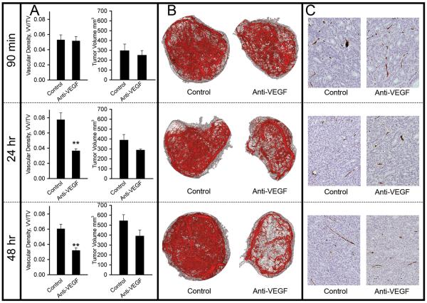
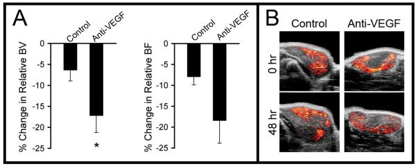
Experimental design: Preclinical studies were done using ex vivo microcomputed tomography and in vivo ultrasound imaging to characterize tumor vasculature in a human HM-7 colorectal xenograft model treated with the anti-VEGF antibody G6-31. Clinical evaluation was by quantitative magnetic resonance imaging in 10 patients with metastatic colorectal cancer treated with bevacizumab.
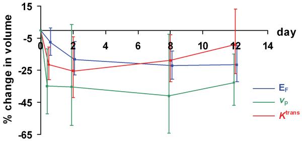
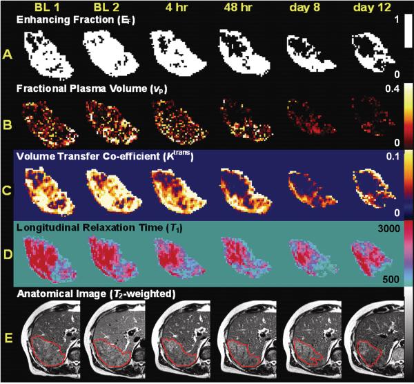
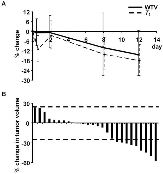
Results: Microcomputed tomography experiments showed reduction in perfused vessels within 24 to 48 h of G6-31 drug administration (P <or= 0.005). Ultrasound imaging confirmed reduced tumor blood volume within the same time frame (P = 0.048). Consistent with the preclinical results, reductions in enhancing fraction and fractional plasma volume were detected in patient colorectal cancer metastases within 48 h after a single dose of bevacizumab that persisted throughout one cycle of therapy. These effects were followed by resolution of edema (P = 0.0023) and tumor shrinkage in 9 of 26 tumors at day 12.
Conclusion: These data suggest that VEGF-specific inhibition induces rapid structural and functional effects with downstream significant antitumor activity within one cycle of therapy. This finding has important implications for the design of early-phase clinical trials that incorporate physiologic imaging. The study shows how animal data help interpret clinical imaging data, an important step toward the validation of image biomarkers of tumor structure and function.
Figures
Comment in
-
Has quantitative multimodal imaging of treatment response arrived?Clin Cancer Res. 2009 Nov 1;15(21):6473-5. doi: 10.1158/1078-0432.CCR-09-2257. Epub 2009 Oct 27. Clin Cancer Res. 2009. PMID: 19861463 Free PMC article.
References
-
- Senger DR, Galli SJ, Dvorak AM, et al. Tumor cells secrete a vascular permeability factor that promotes accumulation of ascites fluid. Science. 1983;219:983–985. - PubMed
-
- Leung DW, Cachianes G, Kuang WJ, Goeddel DV, Ferrara N. Vascular endothelial growth factor is a secreted angiogenic mitogen. Science. 1989;246:1306–1309. - PubMed
-
- Ferrara N, Gerber HP, LeCouter J. The biology of VEGF and its receptors. Nat Med. 2003;9:669–676. - PubMed
-
- Kerr DJ. Targeting angiogenesis in cancer: clinical development of bevacizumab. Nat Clin Pract Oncol. 2004;1:39–43. - PubMed
-
- Hurwitz H, Fehrenbacher L, Novotny W, et al. Bevacizumab plus irinotecan, fluorouracil, and leucovorin for metastatic colorectal cancer. N Engl J Med. 2004;350:2335–2342. - PubMed
Publication types
MeSH terms
Substances
Grants and funding
LinkOut - more resources
Full Text Sources
Other Literature Sources
Medical
Research Materials
Miscellaneous
