

Comment
doi: 10.1158/1078-0432.CCR-09-2257.
Epub 2009 Oct 27.
Has quantitative multimodal imaging of treatment response arrived?
Affiliations
- PMID: 19861463
- PMCID: PMC2783451
- DOI: 10.1158/1078-0432.CCR-09-2257
Item in Clipboard
Comment
Has quantitative multimodal imaging of treatment response arrived?
Clin Cancer Res.
.
Abstract
Although there have been dramatic increases in the range and quality of information available from noninvasive imaging methods, their application in clinical trials has been limited. One promising approach is to apply imaging techniques in preclinical studies designed to mimic a corresponding clinical trial in order to inform that trial.
Figures
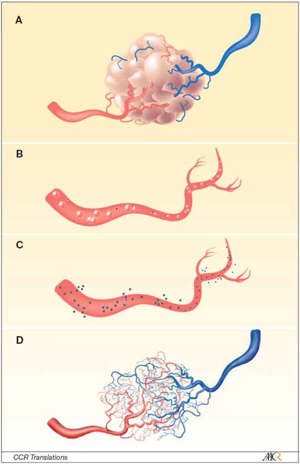
The Figure displays a cartoon depiction of methods to characterize the distinctively fragile and leaky blood vessels associated with tumors (panel a). In the O’Connor et al study, the authors made use of in vivo dynamic contrast enhanced magnetic resonance imaging (DCE-MRI) and microbubble contrast enhanced sonography (MCES), as well as ex vivo micro computed tomography (μCT). The contrast agents they selected for their study allowed them to probe different aspects of the tumor vasculature. For example, since the contrast agent used in MCES is intravascular (panel b), it can report on relative blood flow (rBF), relative blood volume (rBV), and mean transit time (MTT), whereas the DCE-MRI contrast agent is an extravasacular agent and therefore reports on plasma volume (vp), extravascular extracellular volume fraction (ve), and a mixed measure of vessel perfusion and permeability (Ktrans, panel c). The μCT used an intravascular contrast agent that, when combined with the high-resolution ex vivo imaging, can display the vessels in 3D (panel d) and determine vascular volume. As these methods report on different, but related, aspects of tumor vessels, it is reasonable to hypothesize that more can be learned about, for example, a drug’s mechanism of action by studying all three. The O’Connor et al paper investigates the changes these techniques report during a longitudinal study of treatment response.
Comment on
-
Quantifying antivascular effects of monoclonal antibodies to vascular endothelial growth factor: insights from imaging.Clin Cancer Res. 2009 Nov 1;15(21):6674-82. doi: 10.1158/1078-0432.CCR-09-0731. Epub 2009 Oct 27. Clin Cancer Res. 2009. PMID: 19861458 Free PMC article.
References
-
- Eisenhauer EA, Therasse P, Bogaerts J, et al. New response evaluation criteria in solid tumours: revised RECIST guideline (version 1.1) Eur J Cancer. 2009;45:228–47. - PubMed
Publication types
MeSH terms
Grants and funding
LinkOut - more resources
Full Text Sources
Medical
