Multicenter Study
. 2010 Jan 14;115(2):379-87.
doi: 10.1182/blood-2009-05-221549. Epub 2009 Oct 27.

Association of factor H autoantibodies with deletions of CFHR1, CFHR3, CFHR4, and with mutations in CFH, CFI, CD46, and C3 in patients with atypical hemolytic uremic syndrome

Affiliations
Multicenter Study

Association of factor H autoantibodies with deletions of CFHR1, CFHR3, CFHR4, and with mutations in CFH, CFI, CD46, and C3 in patients with atypical hemolytic uremic syndrome

Iain Moore et al. Blood. .

Abstract

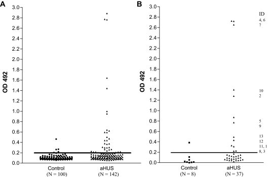
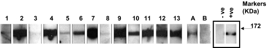
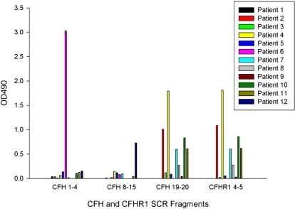
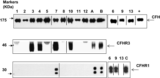
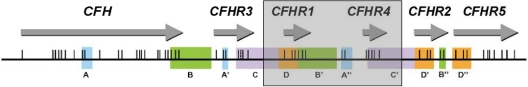
Factor H autoantibodies have been reported in approximately 10% of patients with atypical hemolytic uremic syndrome (aHUS) and are associated with deficiency of factor H-related proteins 1 and 3. In this study we examined the prevalence of factor H autoantibodies in the Newcastle cohort of aHUS patients, determined whether the presence of such autoantibodies is always associated with deficiency of factor H-related proteins 1 and 3, and examined whether such patients have additional susceptibility factors and/or mutations in the genes encoding complement regulator/activators. We screened 142 patients with aHUS and found factor H autoantibodies in 13 individuals (age 1-11 years). The presence of the autoantibodies was confirmed by Western blotting. By using multiplex ligation-dependent probe amplification we measured complement factor H-related (CFHR)1 and CFHR3 copy number. In 10 of the 13 patients there were 0 copies of CFHR1, and in 3 patients there were 2. In 3 of the patients with 0 copies of CFHR1 there was 1 copy of CFHR3, and these individuals exhibited a novel deletion incorporating CFHR1 and CFHR4. In 5 patients mutations were identified: 1 in CFH, 1 in CFI, 1 in CD46, and 2 in C3. The latter observation emphasizes that multiple concurrent factors may be necessary in individual patients for disease manifestation.


Figures

Figure 1
Figure 1
Detection of factor H autoantibodies. Factor H autoantibodies in aHUS patients and control subjects were detected by the use of a sensitive ELISA as described in “Factor H autoantibody assay.” (A) OD492 for 100 control subjects and 142 aHUS patients. (B) OD492 for 8 control subjects and 37 aHUS patients with an uncorrected OD value greater than or close to 0.215 (the mean + 2SD for the 100 control subjects) after background reactivity to BSA has been subtracted to exclude false-positive interactions. The identity of the 13 factor H autoantibody–positive patients is shown.
Figure 2
Figure 2
Analysis of antifactor H binding by Western blotting. Purified factor H was run out on 10% SDS-PAGE and transferred to nitrocellulose. Strips of nitrocellulose were then incubated with sera collected from subjects and bound antibody detected as described in “Methods.” ECL Western blotting substrate was used to visualize bound antibody. The same secondary reagent and exposure times were used throughout. Sera from known factor H autoantibody–positive and –negative subjects were used to allow standardization across multiple experiments. The positive (+ve) and negative controls (−ve) are on adjacent strips on the same autorad film and are shown as representative control signals (black box). The factor H autoantibody patients (1-13) and 2 normal subjects, A and B, are shown. These data are from a collection of sequential experiments. Molecular weight markers are shown, and the data are representative of at least 3 independent experiments.
Figure 3
Figure 3
Autoantibody reactivity with short factor H fragments. Autoantibody binding to factor H fragments (corresponding to SCRs 1-4, 8-15, and 19-20) and a factor H–related protein 1 fragment (SCR 4-5) was assessed by the use of ELISA in a similar manner to the original autoantibody screen. Molar equivalent concentrations of the SCR fragments were coated onto separate ELISA plates, and a BSA subtraction was performed. Results are representative of 3 separate experiments.
Figure 4
Figure 4
Analysis of factor H, factor H–related protein 3, and factor H–related protein 1 in patients with factor H autoantibodies. Sera from subjects were run out on 10% SDS-PAGE and transferred to nitrocellulose. Factor H, factor H–related protein 3, and factor H–related protein 1 were then detected by staining as described in the Methods section. ECL Western blotting substrate was used to visualize bound antibody. Sera was available from all 13 patients (samples 1-13) for analysis of factor H and factor H–related protein 1, but only from 9 patients for factor H–related protein 3. These 9 samples plus control samples were run on parallel gels. A, B, and C are normal controls known to have 2 copies of CFHR1 and CFHR3. Data from the remaining 3 samples are shown on the right. Purified factor H (equivalent to 0.5 mg/mL) was used as a positive control (+) in the smaller antifactor H blot. Black vertical lines indicate a repositioned gel lane, and black boxes illustrate the individual blots used. This figure is representative of several independent experiments.
Figure 5
Figure 5
A novel deletion including CFHR1 and CFHR4. Position of the genes encoding factor H and the factor H–related proteins in a centromeric segment of the RCA cluster at 1q32. Regions of high sequence identity (originally determined by Male et al) are indicated by the same letter and color. Exons are indicated as vertical lines. The shaded box shows the presence of a novel ∼ 125-kb deletion, which includes CFHR1 and CFHR4. This deletion occurs within the duplicons C/C′ as a result of nonallelic homologous recombination.

Comment in

  • aHUS: a disorder with many risk factors.
    Rodríguez de Córdoba S. Rodríguez de Córdoba S. Blood. 2010 Jan 14;115(2):158-60. doi: 10.1182/blood-2009-11-252627. Blood. 2010. PMID: 20075170 No abstract available.

References

    1. Kavanagh D, Goodship THJ, Richards A. Atypical haemolytic uraemic syndrome. Br Med Bull. 2006;77–78(1):5–22. - PubMed
    1. Richards A, Buddles MR, Donne RL, et al. Factor H mutations in hemolytic uremic syndrome cluster in exons 18-20, a domain important for host cell recognition. Am J Hum Genet. 2001;68(2):485–490. - PMC - PubMed
    1. Kavanagh D, Kemp EJ, Mayland E, et al. Mutations in complement factor I predispose to development of atypical hemolytic uremic syndrome. J Am Soc Nephrol. 2005;16(7):2150–2155. - PubMed
    1. Richards A, Kemp EJ, Liszewski MK, et al. Mutations in human complement regulator, membrane cofactor protein (CD46), predispose to development of familial hemolytic uremic syndrome. Proc Natl Acad Sci U S A. 2003;100(22):12966–12971. - PMC - PubMed
    1. Goicoechea de Jorge E, Harris CL, Esparza-Gordillo J, et al. Gain-of-function mutations in complement factor B are associated with atypical hemolytic uremic syndrome. Proc Natl Acad Sci U S A. 2007;104(1):240–245. - PMC - PubMed

Publication types

MeSH terms