. 2010 Jan 1;285(1):723-30.
doi: 10.1074/jbc.M109.033829. Epub 2009 Oct 27.

Crystal structure of glucagon-like peptide-1 in complex with the extracellular domain of the glucagon-like peptide-1 receptor

Affiliations

Crystal structure of glucagon-like peptide-1 in complex with the extracellular domain of the glucagon-like peptide-1 receptor

Christina Rye Underwood et al. J Biol Chem. .

Abstract

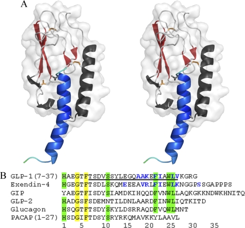
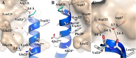
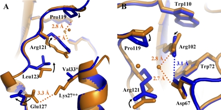
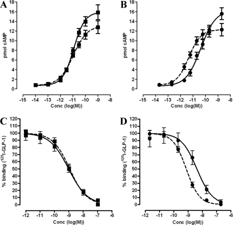
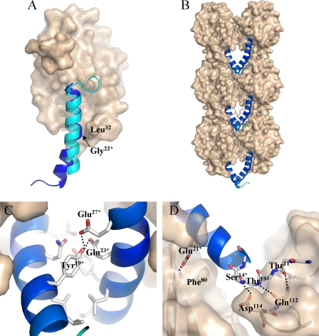
GLP-1 (glucagon-like peptide-1) is an incretin released from intestinal L-cells in response to food intake. Activation of the GLP-1 receptor potentiates the synthesis and release of insulin from pancreatic beta-cells in a glucose-dependent manner. The GLP-1 receptor belongs to class B of the G-protein-coupled receptors, a subfamily characterized by a large N-terminal extracellular ligand binding domain. Exendin-4 and GLP-1 are 50% identical, and exendin-4 is a full agonist with similar affinity and potency for the GLP-1 receptor. We recently solved the crystal structure of the GLP-1 receptor extracellular domain in complex with the competitive antagonist exendin-4(9-39). Interestingly, the isolated extracellular domain binds exendin-4 with much higher affinity than the endogenous agonist GLP-1. Here, we have solved the crystal structure of the extracellular domain in complex with GLP-1 to 2.1 Aresolution. The structure shows that important hydrophobic ligand-receptor interactions are conserved in agonist- and antagonist-bound forms of the extracellular domain, but certain residues in the ligand-binding site adopt a GLP-1-specific conformation. GLP-1 is a kinked but continuous alpha-helix from Thr(13) to Val(33) when bound to the extracellular domain. We supplemented the crystal structure with site-directed mutagenesis to link the structural information of the isolated extracellular domain with the binding properties of the full-length receptor. The data support the existence of differences in the binding modes of GLP-1 and exendin-4 on the full-length GLP-1 receptor.


Figures

FIGURE 1.
FIGURE 1.
Structure of the GLP-1-bound ECD of the GLP-1R. A, stereoview of GLP-1 (blue) bound to the ECD of the GLP-1R (α-helix in black, β-strands in red, and loops in gray). Disulfide bridges are shown as orange sticks. Residues Cys62–Asp671) and Ala70–Gly752) constitute the first region of antiparallel β-sheets, and the second region is comprised of residues Gly78–Ser843) and His99–Thr1054), which is shown in red. Our final structure contains GLP-1 residues Gly10*–Gly35*. The residues that interact with GLP-1R ECD lie within Ala24* and Val33*, which are shown as sticks. B, sequence alignment of GLP-1, exendin-4, GIP, GLP-2, glucagon, and PACAP(1–27). Fully conserved residues are highlighted in yellow, and partially conserved residues are highlighted in green. The residues of GLP-1 and exendin-4 that interact with GLP-1R ECD are colored blue. The underlined residues symbolize residues of GLP-1 in α-helical conformation when bound to the ECD. Residue number 1 of exendin-4 corresponds to residue number 7 of GLP-1.
FIGURE 2.
FIGURE 2.
Interactions between GLP-1 and GLP-1R ECD. A, ribbon diagram of GLP-1 and its hydrophilic interactions with GLP-1R ECD. GLP-1 is colored in cyan, and residues Gln23*, Lys26*, Glu27*, Trp31*, and Val33* are illustrated as sticks. Receptor residues Arg121, Leu123, Glu127, and Glu128 are shown as sticks. The surface of the hydrophilic binding cavity of ECD is illustrated in gray. B, ribbon diagram of GLP-1 and its hydrophobic interactions with GLP-1R ECD. GLP-1 residues Ala24*, Glu27*, Phe28*, Trp31*, and Leu32* are illustrated as sticks, and so are ECD residues Leu32, Trp39, Asp67, and Arg121. The surface of the hydrophobic binding cavity of ECD is illustrated in gray. C, ribbon diagram illustrating a common motif found in the GLP-1R ECD and in the GIP receptor ECD. The side chain of Arg121 interacts with the backbone carbonyls of Asp67 and Leu32* through a water molecule. GLP-1 residues Leu32* and Val33* are illustrated as sticks, and so are ECD residues Asp67 and Arg121.
FIGURE 3.
FIGURE 3.
Differences between the GLP-1- and exendin-4(9–39)-bound structure of ECD. Ribbon diagrams showing significant differences in side chain conformations between the GLP-1-bound structure and the exendin-4(9–39)-bound structure of GLP-1R ECD. Receptor and ligand residues are highlighted in blue for the GLP-1-bound structure and in orange for the exendin-4(9–39)-bound structure. Water molecules in orange are present only in the exendin-4(9–39)-bound structure. A, one diverging residue, Val33* of GLP-1 and Lys27** of exendin-4(9–39), causes a shift in the conformations of four residues, namely Glu127, Leu123, Arg121, and Pro119. B, GLP-1-specific conformations affect the conserved core of the ECD by rotating the guanidine group of Arg102 and by decreasing the distance between Asp67 and Arg102 compared with the exendin-4(9–39)-bound structure without affecting the relative position of Trp72 and Trp110.
FIGURE 4.
FIGURE 4.
Functional and binding properties of the L32A GLP-1R mutant. Upper panel, stimulation of cAMP production by transiently transfected HEK293 cells expressing the L32A mutant by GLP-1 (squares, A) and exendin-4 (circles, B). Dashed dose-response curves represent cAMP production by GLP-1 and exendin-4 at the wild-type GLP-1R, respectively. Lower panel, competition binding assay on plasma membranes from transiently transfected HEK293 cells expressing the L32A mutant. GLP-1 binding curves are presented with squares (C) and exendin-4 curves with circles (D). Dashed binding curves represent 125I-GLP-1 displacement by GLP-1 and exendin-4 at the wild-type GLP-1R. Data are normalized according to 125I-GLP-1 binding and correspond to three independent experiments performed in duplicate.
FIGURE 5.
FIGURE 5.
Crystal packing. A, superposition of ECD-bound GLP-1 (blue) and exendin-4(9–39) (cyan). GLP-1 residue Gly22* denotes a kink in the α-helix, which is situated in close proximity to Leu32 of the ECD. B, crystal packing involving symmetry-related complex molecules. C, ribbon diagram of GLP-1 (blue) and its interactions with the ligand of a symmetry-related molecule. Residues Tyr19*, Gln23*, and Glu27* are shown as sticks, and the surface of the GLP-1R ECD is shown in gray. The packing of complex molecules allows Tyr19* to interact with Gln23* (3 Å) and Glu27* (2.5 Å) in a symmetry-related ligand molecule. D, interactions between GLP-1 and residues of symmetry-related ECD molecules. GLP-1 residues Thr11*, Thr13*, Ser14*, and Glu21* are shown as sticks. The backbone carbonyl of Thr11* could form a weak hydrogen bond (3.2 Å) to the backbone amide of Gln112, the backbone amide of Thr13* could form a hydrogen bond (2.9 Å) to the backbone carbonyl of Gln112, and the backbone amide of Ser14* may form a hydrogen bond (3 Å) to the side chain of Asp114. The side chain of Glu21* forms a hydrogen bond to the backbone amide of Phe80 (2.6 Å).

Similar articles

Cited by

References

    1. Mojsov S., Heinrich G., Wilson I. B., Ravazzola M., Orci L., Habener J. F. (1986) J. Biol. Chem. 261, 11880–11889 - PubMed
    1. Schmidt W. E., Siegel E. G., Creutzfeldt W. (1985) Diabetologia 28, 704–707 - PubMed
    1. Flint A., Raben A., Astrup A., Holst J. J. (1998) J. Clin. Invest. 101, 515–520 - PMC - PubMed
    1. Orskov C., Holst J. J., Nielsen O. V. (1988) Endocrinology 123, 2009–2013 - PubMed
    1. Wettergren A., Schjoldager B., Mortensen P. E., Petersen H., Ørskov C., Holst J. J. (1993) Digestion 54, 384–385

Publication types

MeSH terms

Associated data