

Case Reports
doi: 10.4103/0301-4738.57160.
Amyloidosis of lacrimal gland
Affiliations
- PMID: 19861750
- PMCID: PMC2812767
- DOI: 10.4103/0301-4738.57160
Item in Clipboard
Case Reports
Amyloidosis of lacrimal gland
Indian J Ophthalmol.
2009 Nov-Dec.
Abstract
Primary localized amyloidosis of lacrimal gland is a rare occurrence. This report describes a female patient with isolated amyloidosis of the lacrimal gland. A 45-year-old Indian woman presented with a swelling over the left lacrimal gland region. Computed tomography showed uniform enlargement of the lacrimal gland. A lacrimal gland biopsy revealed amyloidosis. No systemic involvement was detected on further investigation. To our knowledge, this is the first report of lacrimal gland amyloidosis from India and our report also highlights the importance of lacrimal gland biopsy in diagnosing lacrimal gland masses.
Figures

Clinical photograph showing left ptosis and inferior globe displacement
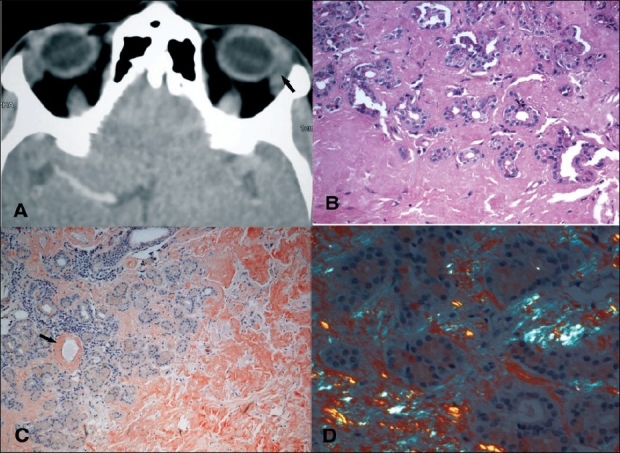
(A) Computed tomography scan (axial view) showing welldefined enlargement of left lacrimal gland (arrow), (B) Photomicrograph of lacrimal gland biopsy illustrating acini infiltrated with amorphous eosinophilic material which has completely replaced the acini in certain areas (H & E, ×100), (C) Section stained with Congo red showing brick-red staining of the amorphous material consistent with amyloid replacing the entire lobule on the right side and deposited in the perivascular region (arrow) (Congo red stain, original magnification ×200), (D) Section stained with Congo red and viewed under polarized light illustrates the apple-green birefringence characteristic of amyloidosis
Comment in
-
Amyloidosis of lacrimal gland.Indian J Ophthalmol. 2010 Jul-Aug;58(4):348. doi: 10.4103/0301-4738.64137. Indian J Ophthalmol. 2010. PMID: 20534938 Free PMC article. No abstract available.
Similar articles
-
Lacrimal gland amyloidosis: a clinicopathological correlation of a rare disorder and review of literature.Ocul Immunol Inflamm. 2014 Aug;22(4):300-5. doi: 10.3109/09273948.2013.850100. Epub 2013 Nov 8. Ocul Immunol Inflamm. 2014. PMID: 24205967 Review.
-
Primary localized orbital amyloidosis.Ann Ophthalmol. 1986 May;18(5):165-7. Ann Ophthalmol. 1986. PMID: 3717836
-
Lacrimal gland amyloidosis.Ophthalmic Plast Reconstr Surg. 2006 Jul-Aug;22(4):306-8. doi: 10.1097/01.iop.0000222354.44000.eb. Ophthalmic Plast Reconstr Surg. 2006. PMID: 16855510
-
Clinical and computed tomographic characteristics of amyloid tumor of the lacrimal gland.Ophthalmology. 1996 Aug;103(8):1233-6. doi: 10.1016/s0161-6420(96)30517-4. Ophthalmology. 1996. PMID: 8764792 Review.
-
Isolated bilateral amyloidosis of the lacrimal gland: Case report from a referral center in Mexico.J Fr Ophtalmol. 2020 Jun;43(6):e207-e209. doi: 10.1016/j.jfo.2019.10.008. Epub 2020 Apr 29. J Fr Ophtalmol. 2020. PMID: 32360080 No abstract available.
Cited by
-
Primary Isolated Lacrimal Gland Amyloidosis: A Case Report and Review of the Literature.Cureus. 2019 Nov 29;11(11):e6258. doi: 10.7759/cureus.6258. Cureus. 2019. PMID: 31893184 Free PMC article.
-
Lacrimal Gland Amyloidosis in an Elderly Patient.Case Rep Ophthalmol. 2020 Mar 6;11(1):100-105. doi: 10.1159/000505480. eCollection 2020 Jan-Apr. Case Rep Ophthalmol. 2020. PMID: 32308610 Free PMC article.
-
Amyloidosis of lacrimal gland.Indian J Ophthalmol. 2010 Jul-Aug;58(4):348. doi: 10.4103/0301-4738.64137. Indian J Ophthalmol. 2010. PMID: 20534938 Free PMC article. No abstract available.
-
Amyloidosis of lacrimal gland: authors' reply.Indian J Ophthalmol. 2010 Sep-Oct;58(5):450-1. doi: 10.4103/0301-4738.67059. Indian J Ophthalmol. 2010. PMID: 20689214 Free PMC article. No abstract available.
-
Ocular adnexal and orbital amyloidosis: a case series and literature review.Int Ophthalmol. 2016 Apr;36(2):281-98. doi: 10.1007/s10792-015-0138-7. Epub 2015 Oct 14. Int Ophthalmol. 2016. PMID: 26466598 Review.
References
-
- Leibovitch I, Selva D, Goldberg RA, Sullivan TJ, Saeed P, Davis G, et al. Periocular and orbital amyloidosis. Clinical charateristics, management and outcome. Ophthalmology. 2006;113:1657–64. - PubMed
-
- Chuan Cheng JY, Fong KS, Cheah ES. Lacrimal gland amyloidosis. Ophthal Plast Reconstr Surg. 2006;22:306–8. - PubMed
-
- Knowles DM, Jakobiec FA, Rosen M, Howard G. Amyloidosis of the orbit and adnexae. Surv Opthalmol. 1975;19:367–84. - PubMed
-
- Conlon MR, Chapman WB, Burt WL, Larocque BJ, Hearn SA. Primary localized amyloidosis of the lacrimal glands. Ophthalmology. 1991;98:1556–9. - PubMed
Publication types
MeSH terms
LinkOut - more resources
Full Text Sources
Medical
