

A rare cause of nasolacrimal duct obstruction: dentigerous cyst in the maxillary sinus
- PMID: 19861752
- PMCID: PMC2812769
- DOI: 10.4103/0301-4738.57161
A rare cause of nasolacrimal duct obstruction: dentigerous cyst in the maxillary sinus
Abstract
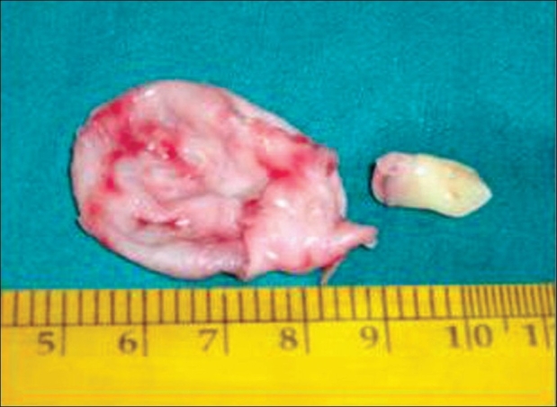
The most common abnormality of the lacrimal drainage system is congenital or acquired nasolacrimal duct obstruction. The causes of acquired nasolacrimal duct obstruction may be primary or secondary. The secondary acquired obstructions may result from infection, inflammation, neoplasm, trauma or mechanical causes. The maxillary sinus cysts usually obstruct the nasolacrimal duct mechanically. Dentigerous cysts are one of the main types of maxillary cysts. These cysts are benign odontogenic cysts which are associated with the crowns of unerupted teeth. The clinical documentations of mechanical nasolacrimal duct obstructions due to a dentigerous cyst in the maxillary sinus are very rare in literature. In this case report, we describe a dentigerous cyst with a supernumerary tooth in the maxillary sinus in an 11-year-old male child causing an obstruction to the nasolacrimal duct. The case was successfully managed surgically by Caldwell Luc approach.
Figures


References
-
- Bartley GB. Acquired lacrimal drainage obstruction: An etiologic classification system, case reports, and a review of the literature. Ophthal Plast Reconstr Surg. 1992;8:237–49. - PubMed
-
- Alexandrakis G, Hubbell RN, Aitken PA. Nasolacrimal duct obstruction secondary to ectopic teeth. Ophthalmology. 2000;107:189–92. - PubMed
-
- Bajaj MS, Mahindrakar A, Pushker N. Dentigerous cyst in the maxillary sinus: A rare cause of nasolacrimal obstruction. Orbit. 2003;22:289–92. - PubMed
-
- Micozkadioglu SD, Erkan AN. Endoscopic removal of a maxillary dentigerous cyst. B-ENT. 2007;3:213–6. - PubMed
-
- Motamedi MHK, Talesh KT. Management of extensive dentigerous cysts. British Dental Journal. 2005;198:203–6. - PubMed
Publication types
MeSH terms
LinkOut - more resources
Full Text Sources
