TGF-beta-induced interleukin-6 participates in transdifferentiation of human Tenon's fibroblasts to myofibroblasts
- PMID: 19862334
- PMCID: PMC2765236
TGF-beta-induced interleukin-6 participates in transdifferentiation of human Tenon's fibroblasts to myofibroblasts
Abstract
Purpose: To gain a better understanding of the roles of interleukins (ILs) in subconjunctival fibrosis, we investigated their expression in transforming growth factor-beta1 (TGF-beta1)-stimulated Tenon's fibroblasts and examined their association with the transdifferentiation of fibroblasts to myofibroblasts.
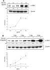
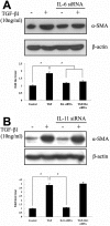
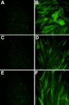
Methods: After primary culture, fibroblasts derived from human Tenon's capsule were exposed to TGF-beta1. The expression of alpha-smooth muscle actin (alpha-SMA) protein was assessed by western immunoblots and immunofluorescence. The mRNA levels of various ILs were also evaluated by multiplex reverse transcription (RT)-PCR. Using the small interfering RNAs (siRNAs) specific for IL-6 and IL-11 and the promoter deletion assay, the contributions of IL-6 and IL-11 to TGF-beta1-induced induction of alpha-SMA were determined.
Results: In human Tenon's fibroblasts, TGF-beta1 stimulated the expression of alpha-SMA protein determined by western blot analysis and also increased the mRNA levels of IL-6 and IL-11 determined by multiplex RT-PCR. On the western immunoblots and immunofluorescence, the increased expression of alpha-SMA was attenuated only by the siRNAs specific for IL-6 but not by the siRNAs specific for IL-11. When the activator protein-1 binding sites of the IL-6 promoter region were deleted, the stimulation effects of TGF-beta1 decreased.
Conclusions: Our data show that autocrine IL-6 may participate in the TGF-beta1-induced transdifferentiation of human Tenon's fibroblasts to myofibroblasts, which is known to be an essential step for subconjunctival fibrosis.
Figures


Similar articles
-
Role of heat shock protein 47 in transdifferentiation of human tenon's fibroblasts to myofibroblasts.BMC Ophthalmol. 2012 Sep 11;12:49. doi: 10.1186/1471-2415-12-49. BMC Ophthalmol. 2012. PMID: 22967132 Free PMC article.
-
The role of focal adhesion kinase in the TGF-β-induced myofibroblast transdifferentiation of human Tenon's fibroblasts.Korean J Ophthalmol. 2012 Feb;26(1):45-8. doi: 10.3341/kjo.2012.26.1.45. Epub 2012 Jan 14. Korean J Ophthalmol. 2012. PMID: 22323885 Free PMC article.
-
Nintedanib inhibits TGF-β-induced myofibroblast transdifferentiation in human Tenon's fibroblasts.Mol Vis. 2018 Dec 9;24:789-800. eCollection 2018. Mol Vis. 2018. PMID: 30636861 Free PMC article.
-
Epigallocatechin-3-gallate increases autophagic activity attenuating TGF-β1-induced transformation of human Tenon's fibroblasts.Exp Eye Res. 2021 Mar;204:108447. doi: 10.1016/j.exer.2021.108447. Epub 2021 Jan 16. Exp Eye Res. 2021. PMID: 33465394
-
Sulforaphane inhibits TGF-β-induced fibrogenesis and inflammation in human Tenon's fibroblasts.Mol Vis. 2024 Mar 29;30:200-210. eCollection 2024. Mol Vis. 2024. PMID: 39563680 Free PMC article.
Cited by
-
Stem-cell therapy and platelet-rich plasma in regenerative medicines: A review on pros and cons of the technologies.J Oral Maxillofac Pathol. 2018 Sep-Dec;22(3):367-374. doi: 10.4103/jomfp.JOMFP_93_18. J Oral Maxillofac Pathol. 2018. PMID: 30651682 Free PMC article. Review.
-
Positive Feedback Loop of SNAIL-IL-6 Mediates Myofibroblastic Differentiation Activity in Precancerous Oral Submucous Fibrosis.Cancers (Basel). 2020 Jun 18;12(6):1611. doi: 10.3390/cancers12061611. Cancers (Basel). 2020. PMID: 32570756 Free PMC article.
-
LINC01605 knockdown induces apoptosis in human Tenon's capsule fibroblasts by inhibiting autophagy.Exp Ther Med. 2022 May;23(5):343. doi: 10.3892/etm.2022.11273. Epub 2022 Mar 22. Exp Ther Med. 2022. PMID: 35401799 Free PMC article.
-
Role of heat shock protein 47 in transdifferentiation of human tenon's fibroblasts to myofibroblasts.BMC Ophthalmol. 2012 Sep 11;12:49. doi: 10.1186/1471-2415-12-49. BMC Ophthalmol. 2012. PMID: 22967132 Free PMC article.
-
Interleukin-6 inhibition in the management of non-infectious uveitis and beyond.J Ophthalmic Inflamm Infect. 2019 Sep 16;9(1):17. doi: 10.1186/s12348-019-0182-y. J Ophthalmic Inflamm Infect. 2019. PMID: 31523783 Free PMC article. Review.
References
-
- Skuta GL, Parrish RK., 2nd Wound healing in glaucoma filtering surgery. Surv Ophthalmol. 1987;32:149–70. - PubMed
-
- Dutt JE, Ledoux D, Baer H, Foster CS. Collagen abnormalities in conjunctiva of patients with cicatricial pemphigoid. Cornea. 1996;15:606–11. - PubMed
-
- Razzaque MS, Foster CS, Ahmed AR. Role of collagen-binding heat shock protein 47 and transforming growth factor-beta1 in conjunctival scarring in ocular cicatricial pemphigoid. Invest Ophthalmol Vis Sci. 2003;44:1616–21. - PubMed
-
- Esson DW, Neelakantan A, Iyer SA, Blalock TD, Balasubramanian L, Grotendorst GR, Schultz GS, Sherwood MB. Expression of connective tissue growth factor after glaucoma filtration surgery in a rabbit model. Invest Ophthalmol Vis Sci. 2004;45:485–91. - PubMed
-
- Andreev K, Zenkel M, Kruse F, Jünemann A, Schlötzer-Schrehardt U. Expression of bone morphogenetic proteins (BMPs), their receptors, and activins in normal and scarred conjunctiva: role of BMP-6 and activin-A in conjunctival scarring? Exp Eye Res. 2006;83:1162–70. - PubMed
Publication types
MeSH terms
Substances
LinkOut - more resources
Full Text Sources
Research Materials
