

Reconstruction of a human hemicornea through natural scaffolds compatible with the growth of corneal epithelial stem cells and stromal keratocytes
- PMID: 19862337
- PMCID: PMC2765239
Reconstruction of a human hemicornea through natural scaffolds compatible with the growth of corneal epithelial stem cells and stromal keratocytes
Abstract
Purpose: To reconstruct a human hemicornea in vitro by means of limbal stem cells cultured onto human keratoplasty lenticules (HKLs) and to obtain a natural corneal graft for clinical applications.
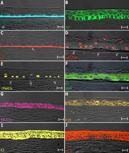
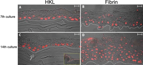
Methods: Limbal stem cells were seeded onto HKLs with or without the presence of feeder layers of lethally irradiated 3T3-J2 cells and compared with the current "gold standard" scaffold, i.e., the fibrin glue. The effects of the scaffold on the preservation of stemness and/or induction of differentiation pathways were investigated through analysis of a variety of markers, including p63 and DeltaNp63alpha for stemness, 14-3-3sigma for early differentiation, keratins 3, 14, 12, and 19 to determine cell phenotype, and alpha6, beta1, and beta4 integrins to evaluate interactions with the stroma. Integrity of the stroma was assessed through analysis of keratan sulfate, CD-34 and aldehyde dehydrogenase 3A1 (ALDH3A1) (for keratocytes), visual system homeobox 1 (VSX1), and alpha-smooth muscle actin (alpha-SMA) (for fibroblasts and myofibroblasts). The structural properties of the reconstructed "hemicornea" were investigated through scanning electron microscopy. To evaluate the preservation of the stemness potential, cells were trypsinized from each scaffold and clonogenic/proliferative characteristics analyzed.
Results: Limbal stem cells expanded onto HKLs gave rise to a stratified squamous keratinized epithelium morphologically similar to that of normal corneas. The resulting corneal epithelium was characterized by basal expression of p63 and DeltaNp63alpha, while expression of 14-3-3sigma, keratin 3, and keratin 12 was found in the upper cell layers. The basal cuboidal epithelial cells were anchored to the basement membrane and expressed keratin 14 and alpha6, beta1, and beta4 integrins. In the stroma of HKLs, keratocytes maintained the biosynthetic and phenotypic appearances typical of resting/quiescent cells and expressed keratan sulfate, CD-34, and ALDH3A1. Fibroblastic transformation was observed with the appearance of VSX1 and alpha-SMA. Scanning electron microscopy analysis showed that HKLs maintained their native conformation with collagen fibrils interconnected to the network and parallel to the corneal surface. HKLs did not alter the clonogenic/proliferative capacity of limbal stem cells. No differences were seen when HKL was compared to fibrin glue, one of the scaffolds currently used for limbal stem cell transplantation.
Conclusions: Our findings demonstrate that HKL could be a suitable scaffold for corneal epithelial stem cells as they were shown to proliferate, express differentiation markers, and bind to the underlying stroma with no alterations in clonogenic potential. HKLs have some advantages over currently used scaffolds, such as the possibility to allow cell growth with no feeder layers, to be freeze dried, and to preserve the integrity and viability of stromal keratocytes. The development of a tissue-engineered "hemicornea" might offer new therapeutic perspectives to patients affected by total limbal stem cell deficiency with stromal scarring.
Figures


Similar articles
-
Development of a hemicornea from human primary cell cultures for pharmacotoxicology testing.Cell Biol Toxicol. 2007 Jul;23(4):279-92. doi: 10.1007/s10565-006-0191-0. Epub 2007 Feb 13. Cell Biol Toxicol. 2007. PMID: 17380411
-
Easy xeno-free and feeder-free method for isolating and growing limbal stromal and epithelial stem cells of the human cornea.PLoS One. 2017 Nov 17;12(11):e0188398. doi: 10.1371/journal.pone.0188398. eCollection 2017. PLoS One. 2017. PMID: 29149196 Free PMC article.
-
Development of a rabbit corneal equivalent using an acellular corneal matrix of a porcine substrate.Mol Vis. 2008;14:2180-9. Epub 2008 Nov 30. Mol Vis. 2008. PMID: 19052652 Free PMC article.
-
Identification and characterization of limbal stem cells.Exp Eye Res. 2005 Sep;81(3):247-64. doi: 10.1016/j.exer.2005.02.016. Exp Eye Res. 2005. PMID: 16051216 Review.
-
[Porous matrix and primary-cell culture: a shared concept for skin and cornea tissue engineering].Pathol Biol (Paris). 2009 Jun;57(4):290-8. doi: 10.1016/j.patbio.2008.04.014. Epub 2008 Jul 3. Pathol Biol (Paris). 2009. PMID: 18602223 Review. French.
Cited by
-
Reconstruction of the corneal epithelium with induced marrow mesenchymal stem cells in rats.Mol Vis. 2010 Jul 14;16:1304-16. Mol Vis. 2010. PMID: 20664793 Free PMC article.
-
[Regenerative medicine for the corneal epithelium : Cell therapy from bench to bedside].Ophthalmologie. 2022 Sep;119(9):891-901. doi: 10.1007/s00347-022-01674-8. Epub 2022 Jun 24. Ophthalmologie. 2022. PMID: 35925345 Review. German.
-
[The emerging technology of tissue engineering : Focus on stem cell niche].Ophthalmologe. 2017 Apr;114(4):327-340. doi: 10.1007/s00347-017-0468-0. Ophthalmologe. 2017. PMID: 28243750 Review. German.
-
Pre-Clinical Cell-Based Therapy for Limbal Stem Cell Deficiency.J Funct Biomater. 2015 Aug 28;6(3):863-88. doi: 10.3390/jfb6030863. J Funct Biomater. 2015. PMID: 26343740 Free PMC article. Review.
-
Decellularized human cornea for reconstructing the corneal epithelium and anterior stroma.Tissue Eng Part C Methods. 2012 May;18(5):340-8. doi: 10.1089/ten.TEC.2011.0072. Epub 2011 Dec 22. Tissue Eng Part C Methods. 2012. PMID: 22082039 Free PMC article.
References
-
- Tseng SC, Prabhasawat P, Barton K, Gray T, Meller D. Amniotic membrane transplantation with or without limbal allografts for corneal surface reconstruction in patients with limbal stem cell deficiency. Arch Ophthalmol. 1998;116:431–41. - PubMed
-
- Tsai RJ, Li LM, Chen JK. Reconstruction of damaged corneas by transplantation of autologous limbal epithelial cells. N Engl J Med. 2000;343:86–93. - PubMed
-
- Koizumi N, Inatomi T, Suzuki T, Sotozono C, Kinoshita S. Cultivated corneal epithelial stem cell transplantation in ocular surface disorders. Ophthalmology. 2001;108:1569–74. - PubMed
-
- Mimura T, Amano S, Usui T, Araie M, Ono K, Akihiro H, Yokoo S, Yamagami S. Transplantation of corneas reconstructed with cultured adult human corneal endothelial cells in nude rats. Exp Eye Res. 2004;79:231–7. - PubMed
-
- Amano S, Shimomura N, Kaji Y, Ishii K, Yamagami S, Araie M. Antigenicity of porcine cornea as xenograft. Curr Eye Res. 2003;26:313–8. - PubMed
Publication types
MeSH terms
Substances
LinkOut - more resources
Full Text Sources
Medical
Research Materials