. 2009 Dec 1;48(47):11283-95.
doi: 10.1021/bi901269j.

Structural basis of the aberrant receptor binding properties of hagfish and lamprey insulins

Affiliations
Free PMC article

Structural basis of the aberrant receptor binding properties of hagfish and lamprey insulins

Waseem Sajid et al. Biochemistry. .
Free PMC article

Abstract

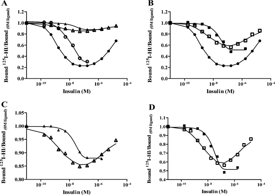
The insulin from the Atlantic hagfish (Myxine glutinosa) has been one of the most studied insulins from both a structural and a biological viewpoint; however, some aspects of its biology remain controversial, and there has been no satisfying structural explanation for its low biological potency. We have re-examined the receptor binding kinetics, as well as the metabolic and mitogenic properties, of this phylogenetically ancient insulin, as well as that from another extant representative of the ancient chordates, the river lamprey (Lampetra fluviatilis). Both insulins share unusual binding kinetics and biological properties with insulin analogues that have single mutations at residues that contribute to the hexamerization surface. We propose and demonstrate by reciprocal amino acid substitutions between hagfish and human insulins that the reduced biological activity of hagfish insulin results from unfavorable substitutions, namely, A10 (Ile to Arg), B4 (Glu to Gly), B13 (Glu to Asn), and B21 (Glu to Val). We likewise suggest that the altered biological activity of lamprey insulin may reflect substitutions at A10 (Ile to Lys), B4 (Glu to Thr), and B17 (Leu to Val). The substitution of Asp at residue B10 in hagfish insulin and of His at residue A8 in both hagfish and lamprey insulins may help compensate for unfavorable changes in other regions of the molecules. The data support the concept that the set of unusual properties of insulins bearing certain mutations in the hexamerization surface may reflect those of the insulins evolutionarily closer to the ancestral insulin gene product.


Figures

Figure 1
Figure 1
Association and dissociation assay. (A and B) Association of 125I-labeled lamprey and hagfish insulin with IM9 cells. The fraction of tracer bound over total is expressed as a function of incubation time. Panel A depicts data from a representative experiment comparing 125I-labeled human insulin (●) and 125I-labeled lamprey insulin (▲), while panel B depicts data from a representative experiment comparing 125I-labeled human insulin (●) and 125I-labeled hagfish insulin (◼). (C) Representative curves of dissociation of 125I-labeled human (●) and 125I-labeled lamprey (▲) insulin in dilution alone (filled symbols) or in the presence of 1 μg/mL human insulin (empty symbols). Bound tracer at time t as a fraction (%) of bound tracer at time zero was plotted as a function of time.
Figure 2
Figure 2
Dose−response curves for ligand-induced acceleration of dissociation. Panel A illustrates dose−response curves for the human insulin tracer in the presence of unlabeled insulin (●), the human insulin tracer in the presence of unlabeled lamprey insulin (○), the lamprey tracer in the presence of unlabeled human insulin (△), and the lamprey tracer in the presence of unlabeled lamprey insulin (▲). Panel B illustrates dose−response curves for the human insulin tracer in the presence of unlabeled insulin (●), the hagfish tracer in the presence of unlabeled human insulin (◻), and the hagfish tracer in the presence of unlabeled hagfish insulin (◼). Panel C is an expansion of the plot of panel A, while panel D is an expansion of the plot of panel B. These expanded scales more clearly demonstrate the presence of reduced negative cooperativity.
Figure 3
Figure 3
Homologous competition for the holo insulin receptor. All curves are plotted as bound:total tracer as a function of the logarithm of the concentration of unlabeled ligand. Competition of 125I-labeled human insulin with increasing concentrations of human insulin (●) is illustrated in the two panels. Competition of 125I-labeled lamprey insulin (▲) is shown in panel A and 125I-labeled hagfish insulin (◼) in Panel B. Data points are averages of duplicate values from a representative experiment.
Figure 4
Figure 4
Functional studies. (A) Dose−response curves for stimulation of lipogenesis in primary rat adipocytes. Lamprey (▲) and hagfish (◼) insulins were compared to human insulin (●) for their ability to stimulate lipogenesis in primary rat adipocytes. Data are averages ± the standard deviation of three experiments performed in triplicate and have been fitted using a four-parameter logistics model. It is plotted as a percentage of the maximum obtained with human insulin as a function of ligand concentration. Panel B illustrates the mitogenic potency by [3H]thymdine incorporation of NIH 3T3 cells overexpressing the insulin receptor. Lamprey (▲) and hagfish (◼) insulins were compared to human insulin (●) in their ability to promote DNA synthesis. Data presented are averages of at least three experiments (performed in triplicate) ± the standard deviation and have been fitted to a four-parameter logistics model. Data are plotted as the percentage of the maximum obtained with 10% FBS as a function of ligand concentration.
Figure 5
Figure 5
Dose−response curves for negative cooperativity (human insulin analogues). Dissociation of prebound 125I-labeled human insulin in the presence of increasing concentrations of human insulin (●) is illustrated in the two curves. Dissociation with increasing concentrations of GlnB4Gly (◼), IleA10Arg (+), and the doubly substituted analogue (▼) (A) and GluB13Asn (◆) and GluB21Val (▲) (B). Curves are illustrated as bound to bound at 0 M cold ligand after dissociation for 30 min. The curves are averages of three assays each made in duplicate. Standard deviations are shown for all the data (smaller than symbol when not visible).
Figure 6
Figure 6
Dose−response curves for negative cooperativity (hagfish insulin analogues). Dissociation of prebound 125I-labeled human insulin in the presence of increasing concentrations of human insulin (●) is illustrated in all three curves. (A) Dissociation with increasing concentrations of HisA8Thr (◻), hagfish (KA9S, KB9S) (○), and AspB10His (△). (B) GlyB4Gln (×), AsnB13Glu (◇), and ValB21Glu (+). (C) Hagfish (KA9S, KB9S) (○) and AspA15Gln (Θ). Curves are illustrated as bound to bound at 0 M cold ligand after dissociation for 30 min. The curves are an average of three assays each conducted in duplicate. Standard deviations are shown for all the data (smaller than the symbol when not visible).
Figure 7
Figure 7
Summary of the contribution of each amino acid. Panel A illustrates the effect of each hagfish insulin substitution in human insulin, and panel B illustrates human insulin substitutions in hagfish insulin. The substitutions marked in red led to a significant decrease in affinity, while the substitutions that led to a significant increase in affinity are marked in green. The blue substitutions had no significant change in affinity. The atomic coordinates [(A) human insulin (Protein Data Bank entry 1G7A) and (B) hagfish insulin (a gift from G. G. Dodson)] were used to construct the diagrams. Molecular graphic images were produced using PyMol from DeLano Scientific LLC.
Figure 8
Figure 8
Simulated dose−response curves for negative cooperativity. The curves were generated using a harmonic oscillator model as previously described (see ref (30) for a detailed explanation). The model contains five binding parameters: a1, rate constant for association with site 1; a2, rate constant for association with site 2; d1, rate constant for dissociation from site 1; d2, rate constant for dissociation from site 2; and kcr, cross-linking constant. For the values of the constants, see ref (30). The simulation shows that for decreasing values of a2, the dose−response curve goes from bell-shaped to sigmoid within the range of ligand concentrations used in the experiments.

References

    1. De Meyts P. (2004) Insulin and its receptor: Structure, function and evolution. BioEssays 26, 1351–1362. - PubMed
    1. Hernandez-Sanchez C.; Mansilla A.; de Pablo F.; Zardoya R. (2008) Evolution of the insulin receptor family and receptor isoform expression in vertebrates. Mol. Biol. Evol. 25, 1043–1053. - PubMed
    1. Wilkinson T. N.; Bathgate R. A. (2007) The evolution of the relaxin peptide family and their receptors. Adv. Exp. Med. Biol. 612, 1–13. - PubMed
    1. Adams T. E.; Epa V. C.; Garrett T. P.; Ward C. W. (2000) Structure and function of the type 1 insulin-like growth factor receptor. Cell. Mol. Life Sci. 57, 1050–1093. - PMC - PubMed
    1. Bathgate R. A.; Ivell R.; Sanborn B. M.; Sherwood O. D.; Summers R. J. (2005) Receptors for relaxin family peptides. Ann. N.Y. Acad. Sci. 1041, 61–76. - PubMed

Publication types