. 2010 Mar;12(3):343-61.
doi: 10.1111/j.1462-5822.2009.01398.x. Epub 2009 Oct 27.

Caenorhabditis is a metazoan host for Legionella

Affiliations

Caenorhabditis is a metazoan host for Legionella

Ann Karen C Brassinga et al. Cell Microbiol. 2010 Mar.

Abstract

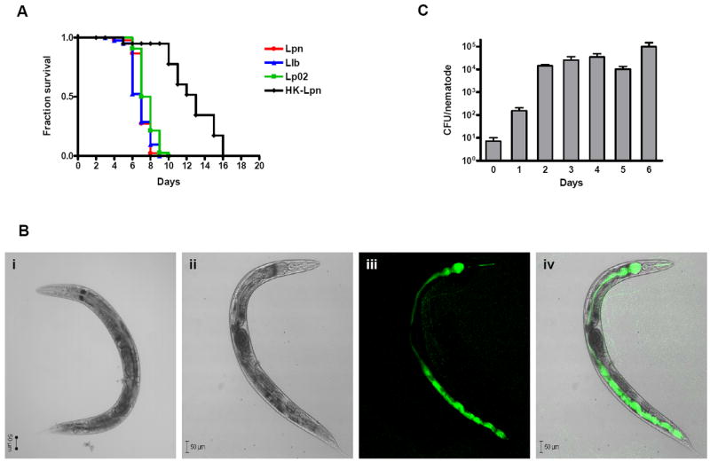
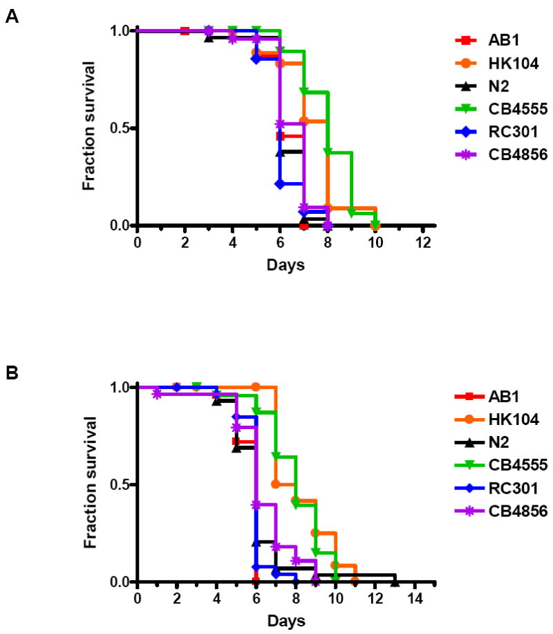
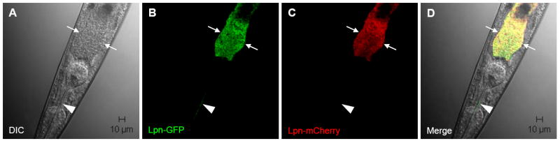
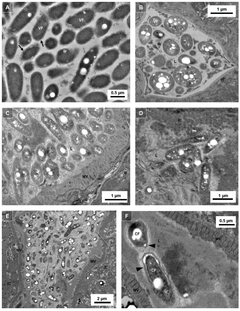
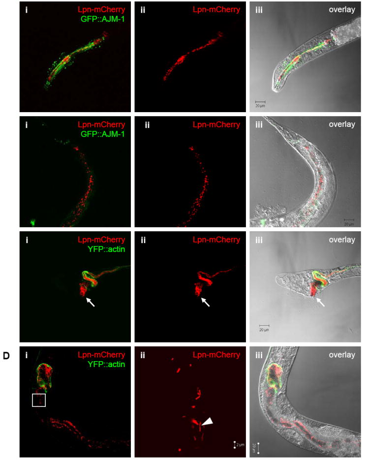
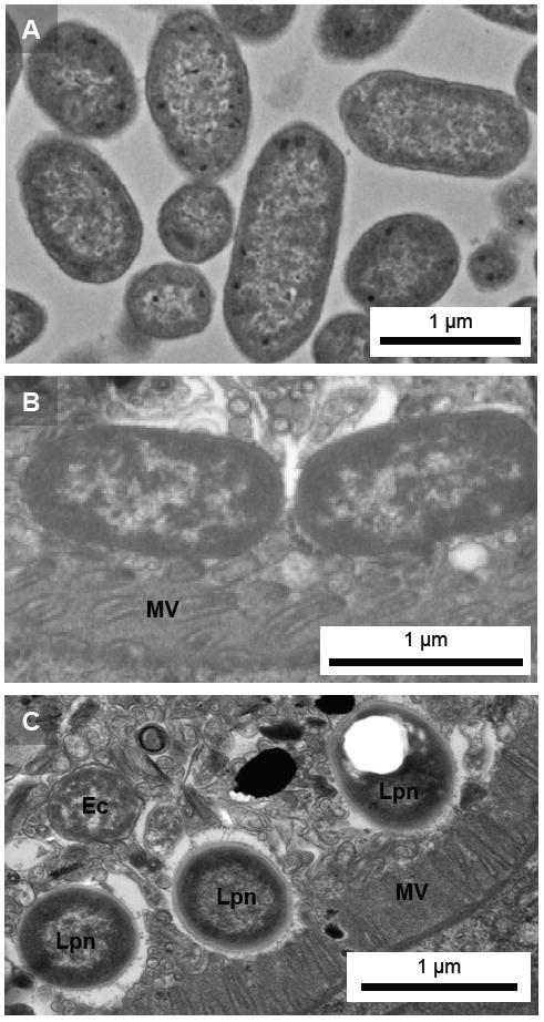
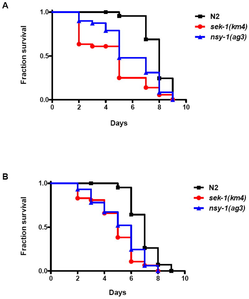
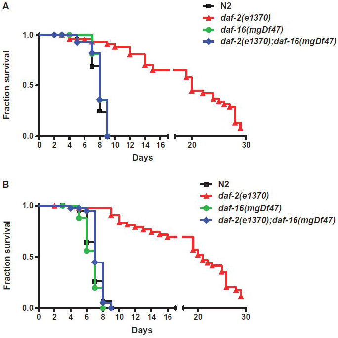
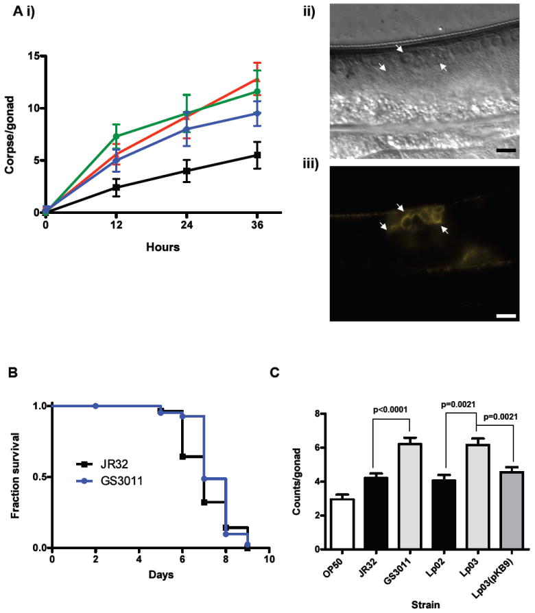
We investigated whether nematodes contribute to the persistence, differentiation and amplification of Legionella species in soil, an emerging source for Legionnaires' disease. Here we show that Legionella spp. colonize the intestinal tracts of Caenorhabditis nematodes leading to worm death. Susceptibility to Legionella is influenced by innate immune responses governed by the p38 mitogen-activated protein kinase and insulin/insulin growth factor-1 receptor signalling pathways. We also show that L. pneumophila colonizes the intestinal tract of nematodes cultivated in soil. To distinguish between transient infection and persistence, plate-fed and soil-extracted nematodes-fed fluorescent strains of L. pneumophila were analysed. Bacteria replicated within the nematode intestinal tract, did not invade surrounding tissue, and were excreted as differentiated forms that were transmitted to offspring. Interestingly, the ultrastructural features of the differentiated bacterial forms were similar to cyst-like forms observed within protozoa, amoeba and mammalian cell lines. While intestinal colonization of L. pneumophila dotA and icmT mutant strains did not alter the survival rate of nematodes in comparison to wild-type strains, nematodes colonized with the dot/icm mutant strains exhibited significantly increased levels of germline apoptosis. Taken together, these studies show that nematodes may serve as natural hosts for these organisms and thereby contribute to their dissemination in the environment and suggest that the remarkable ability of L. pneumophila to subvert host cell signalling and evade mammalian immune responses evolved through the natural selection associated with cycling between protozoan and metazoan hosts.


Figures

Figure 1
Figure 1
Survival of C. elegans fed on Legionella. (A) Kaplan-Meier survival plots of N2 nematodes fed L. pneumophila Philadelphia-1 (Lpn; circles, n=45), L. longbeachae ATCC 33462 (Llb; triangles, n=43), L. pneumophila Philadelphia-1 derivative Lp02 strain (squares; n=44), and heat-killed L. pneumophila Philadelphia-1 (HK-Lpn; diamonds, n=28). Nematode loss was not a significant consideration in the survival assay as the nematodes remained mostly within the bacterial lawn. P < 0.0001 by pairwise comparison by the log-rank test of the each of the strains (Lpn, Llb, Lp02) versus heat-killed. P ≤ 0.01 by pairwise comparison by the log-rank test for each of the strains Lpn and Llb vs. Lp02. Data is representative of one of three independent experiments. (B) Confocal microscopy of Legionella colonization and persistence in wild-type N2 nematodes. DIC images of wild-type N2 nematodes fed (i) heat-killed or (ii-iv) live GFP-expressed L. pneumophila KB130 for five days. (ii) Bright-field image using DIC optics; (iii) KB130 producing GFP colonizing the length of intestinal tract; and (iv) merged image of ii and iii. (C) Bacterial intestinal load of L. pneumophila KB130 within wild-type N2 nematodes (CFU/nematode) over a period of six days. Bacterial loads were determined in triplicate from approximately ten disrupted worms. Data represent mean ± SEM.
Figure 2
Figure 2
Survival of multiple Caenorhabditis nematodes fed Legionella. (A) Kaplan-Meier survival plot of C. elegans AB1 (squares, n=30), C. briggsae HK104 (circles, n=31), C. elegans N2 (triangles, n=30), C. elegans CB4555 (inverted triangles, n=24), C. elegans RC301 (diamonds, n=15), and C. elegans CB4856 (asterisks, n=26) fed L. pneumophila Philadelphia-1 type strain. (B) Kaplan-Meier survival plot of C. elegans AB1 (squares, n=26), C. briggsae HK104 (circles, n=27), C. elegans N2 (triangles, n=29), C. elegans CB4555 (inverted triangles, n=25), C. elegans RC301 (diamonds, n=30), and C. elegans CB4856 (asterisks, n=29) fed L. longbeachae ATCC 33462. P ≤ 0.0002 by pairwise comparison by the log-rank test of each of the nematode strains (HK104 and CB4555) versus wild-type N2 for both Legionella strains. All other nematode strain pairwise comparisons versus wild-type N2 were found to be statistically insignificant. Data is representative of one of three independent experiments.
Figure 3
Figure 3
Confocal microscopy of Legionella colonization and persistence in wild-type N2 nematodes. Wild-type worms were “pulsed” with mCherry-producing KB290 for three days and “chased” with GFP-producing KB130. Representative images after three days of exposure to KB130 are shown. (A) Bright-field image using DIC optics; (B) KB130 producing GPF in the intestinal and pharyngeal lumens; (C) KB290 producing mCherry in the intestinal lumen; and (D) merged images of A, B and C. Arrows demarcate the intestinal tract lumen; arrowhead indicates intact KB130 producing GFP in the pharyngeal lumen.
Figure 4
Figure 4
Intraluminal Legionella exhibit morphologically differentiated forms. Transmission electron microscopic images of (A) Legionella harvested from 6-day old assay plate culture without the presence of N2 nematodes; (B) cross-cut pharynx section of N2 nematodes colonized with L. longbeachae ATCC 33462 (note triangular shape of the grinder); (C) and (D) mid-body cross-cut digestive tract sections of N2 nematodes colonized with L. pneumophila Philadelphia-1 type strain; and (E) and (F) mid-body transverse-cut digestive tract sections of nematodes colonized with L. pneumophila SVir. Note that the thickened cell walls and the white circular spaces within the Legionella bacteria are poly-β-hydroxybutyrate (PHBA) granules. VF, vegetative form; CF, cyst-like form; L, intestinal lumen; IC, intestinal cell; MV, microvilli; arrow, thin cell wall of VF Legionella; arrowhead, thick cell wall of CF Legionella.
Figure 5
Figure 5
Presence of Legionella in nematodes cultivated in a soil environment. Confocal microscopic images of mCherry-producing L. pneumophila KB290 in C. elegans PS3729 (A) head of a L2 stage and (B) tail of a young adult 30 days after initial inoculation, and in WS1904 (C) tail of a L3 stage and (D) mid-body of an adult views 60 days after initial inoculation. Panels i-iii correspond (i) red and green channels showing fluorescent KB290 and nematode intestinal cells, (ii) red channel alone showing only KB290,and (iii) overlayed red channel, green channel, and DIC bright-field images. Green fluorescence indicates the apical tight junctions within the pharynx structure in PS3729 and outlines the intestinal tract in WS1904. Note that image (Dii) is at higher magnification and corresponds to the outlined inset box in (Di). Arrows denote a heavily colonized anus leading to anal swelling; arrowhead demonstrates bacilli that appear to have recently divided.
Figure 6
Figure 6
Ultrastructural analysis of bacteria within C. elegans digestive tract. Transmission electron microscopic images of (A) plate grown E. coli OP50, and transverse-cut sections of the digestive tracts of C. elegans PS3729 nematodes extracted from a soil environment 90 days after initial inoculation with intermittent supplementation of E. coli OP50 detail bacteria embedded within intestinal lumen lining the digestive tract; (B) E. coli OP50, and (C) mixed population of OP50 and Legionella. Note ultrastructural differences between OP50 and Legionella, in particular the thickened cell walls and the white spaces indicating the presence of PHBA in Legionella. Lpn, L. pneumophila; Ec, E. coli OP50; MV, microvilli.
Figure 7
Figure 7
Survival of immunocompromised C. elegans fed Legionella. (A) Kaplan-Meier survival plot of C. elegans N2 (squares, n=45), sek-1(km4) (circles, n=44), and nsy-1(ag3) (triangles, n=45) fed L. pneumophila Philadelphia-1 type strain. (B) Kaplan-Meier survival plot of C. elegans N2 (squares, n=45), sek-1(km4) (circles, n=46), and nsy-1(ag3) (triangles, n=44) fed L. longbeachae type strain. P < 0.0001 by pairwise comparison by the log-rank test of each of the nematode strains [sek-1(km4) and nsy-1(ag3)] versus wild-type N2 for both Legionella strains. Data are representative of one of three independent experiments.
Figure 8
Figure 8
Survival of pathogen and stress-resistant C. elegans fed Legionella. (A) Kaplan-Meier survival plot of C. elegans N2 (squares, n=45), daf-2(e1370) (triangles, n=46), daf-16(mgDf47) (circles, n=46), and daf-2(e1370)/daf-16(mgDf47) (diamonds, n=45) fed L. pneumophila Philadelphia-1 type strain. (B) Kaplan-Meier survival plot of C. elegans N2 (squares, n=45), daf-2(e1370) (triangles, n=45), daf-16(mgDf47) (circles, n=27), and daf-2(e1370)/daf-16(mgDf47) (diamonds, n=41) fed on L. longbeachae type strain. P < 0.0001 by pairwise comparison by the log-rank test of daf-2 versus wild-type N2 for both Legionella strains. All other nematode strain pairwise comparisons versus wild-type N2 were found to be statistically insignificant. Data is representative of one of three independent experiments.
Figure 9
Figure 9
Elevated levels of germline apoptosis in C. elegans. (A) Germline apoptosis in C. elegans fed Legionella: (i) Number of corpse cells counted per gonad (n=10) plotted over time at 0, 12, 24, 36 hr in C. elegans N2 fed on E. coli OP50 (squares), L. pneumophila Philadelphia-1 type strain (triangles), L. longbeachae type strain (circles) and S. Typhimurium SL1344 type strain (diamonds); (ii) DIC image of corpse cells (indicated by arrows) in a gonad of a transgenic C. elegans WS2170 fed L. longbeachae for 36 hr with (iii) corresponding image of fluorescent actin filaments associated with corpse cells. Size marker is 10 μm. (B) Kaplan-Meier survival plot of C. elegans N2 fed L. pneumophila JR32 (squares, n=30) and the isogenic icmT mutant GS3011 (circles, n=44). P > 0.05 by pairwise comparison by the log-rank test. Data representative of three independent experiments. (C) Bar graph of mean corpse cell counts per gonad (n=20) in transgenic C. elegans WS2170 nematodes fed on the designated bacterial strain for 24 hr. P = 0.002 and P = 0.015 by pairwise comparison by the log-rank test of OP50 versus JR32 and Lp02, respectively.

References

    1. Aballay A, Ausubel FM. Programmed cell death mediated by ced-3 and ced-4 protects Caenorhabditis elegans from Salmonella typhimurium-mediated killing. Proc Natl Acad Sci U S A. 2001;98:2735–2739. - PMC - PubMed
    1. Aballay A, Drenkard E, Hilbun LR, Ausubel FM. Caenorhabditis elegans innate immune response triggered by Salmonella enterica requires intact LPS and is mediated by a MAPK signaling pathway. Curr Biol. 2003;13:47–52. - PubMed
    1. Aballay A, Yorgey P, Ausubel FM. Salmonella typhimurium proliferates and establishes a persistent infection in the intestine of Caenorhabditis elegans. Curr Biol. 2000;10:1539–1542. - PubMed
    1. Abu-Zant A, Jones S, Asare R, Suttles J, Price C, Graham J, Kwaik YA. Anti-apoptotic signalling by the Dot/Icm secretion system of L. pneumophila. Cell Microbiol. 2007;9:246–264. - PubMed
    1. Anderson GL, Kenney SJ, Millner PD, Beuchat LR, Williams PL. Shedding of foodborne pathogens by Caenorhabditis elegans in compost-amended and unamended soil. Food Microbiol. 2006;23:146–153. - PubMed

Publication types

MeSH terms

LinkOut - more resources