. 2009 Oct 28:5:14.
doi: 10.1186/1746-4811-5-14.

A simple, flexible and efficient PCR-fusion/Gateway cloning procedure for gene fusion, site-directed mutagenesis, short sequence insertion and domain deletions and swaps

Affiliations

A simple, flexible and efficient PCR-fusion/Gateway cloning procedure for gene fusion, site-directed mutagenesis, short sequence insertion and domain deletions and swaps

Ivan I Atanassov et al. Plant Methods. .

Abstract

Background: The progress and completion of various plant genome sequencing projects has paved the way for diverse functional genomic studies that involve cloning, modification and subsequent expression of target genes. This requires flexible and efficient procedures for generating binary vectors containing: gene fusions, variants from site-directed mutagenesis, addition of protein tags together with domain swaps and deletions. Furthermore, efficient cloning procedures, ideally high throughput, are essential for pyramiding of multiple gene constructs.

Results: Here, we present a simple, flexible and efficient PCR-fusion/Gateway cloning procedure for construction of binary vectors for a range of gene fusions or variants with single or multiple nucleotide substitutions, short sequence insertions, domain deletions and swaps. Results from selected applications of the procedure which include ORF fusion, introduction of Cys>Ser mutations, insertion of StrepII tag sequence and domain swaps for Arabidopsis secondary cell wall AtCesA genes are demonstrated.

Conclusion: The PCR-fusion/Gateway cloning procedure described provides an elegant, simple and efficient solution for a wide range of diverse and complicated cloning tasks. Through streamlined cloning of sets of gene fusions and modification variants into binary vectors for systematic functional studies of gene families, our method allows for efficient utilization of the growing sequence and expression data.


Figures
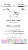
Figure 1
Figure 1
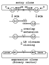
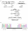
PCR-fusion/Gateway procedure: 'gene fusion' protocol. (A) Schematic of gene fusion protocol. PCR amplification from entry clones (primer pairs F × SR and SF × R) of fragments with short overlapping ends; joining of the PCR fragments by single overlap extension; direct LR clonase II mediated cloning of the assembled DNA fragment into a binary destination vector and generation of binary expression vector (expression clone). The recombination of attL1 and L2 sites with attR1 and R2 sites to give attB sites flanking the fused DNA fragments during the Gateway cloning are designated. Cassette enables efficient selection of entry and expression clones and contains ccdB and chloramphenicol resistance (CmR) genes (indicated with ccd/CmR). (B) Generation of AtCesA7 (aa: 1-1007)/AtCesA8 (aa: 965-985) gene fusion. Two fragments were amplified from the starting entry clones with primers: clone pZ3 (AtCesA7): F(M13for)-gtaaaacgacggccagt × SR- ggagacgaaaggattaattcttacccaaag and clone pZ1(AtCesA8): SF-ctttgggtaagaattaatcctttcgtctcc × R(M13rev)-caggaaacagctatgac. Note that primers F and R are universal M13-forward and M13-reverse primers matching the vector sequence outside the attL1/attL2 region. The sequences of the overlapping ends of the two PCR fragments and gene fusion site are presented. The primer sequences are underline. The AtCesA8 sequences are in bold type. (C) Sequencing of the DNA fusion site in binary expression vector p3K3C1 which contained a AtCesA7 (aa: 1-1007)/AtCesA8 (aa: 965-985) fusion cloned into p3KC binary destination vector. The fusion site and position of SF primer are designated on the DNA sequence.
Figure 2
Figure 2
DNA 'domain deletion' protocol. Schematic representation of a 'domain deletion' using PCR amplification of the two terminal regions of the entry clone adjacent to the DNA domain to be deleted (designated by 'X'). F and R - represent M13 universal primers. SF and SR are complementary PCR primers consisting of two fused sections (a and b) that match regions flanking the deleted domain. The overlap extension results in assembly of the two terminal regions with overlapping ends. The new fragment with deleted DNA domain is directly LR clonase cloned into a binary destination vector producing binary expression vector, (for details see Fig. 1).
Figure 3
Figure 3
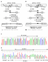
DNA 'domain swap' protocol. (A) Schematic of 'domain swap' protocol. PCR amplification from the domain acceptor entry clone (primer pairs F × SR1 and SF2 × R) of the two terminal regions flanking the DNA domain to be replaced. PCR amplification (primer pair SF1 × SR2) of the donor domain to be inserted. Overlap extension assembly of the three PCR fragments with short overlapping ends (for details see Fig. 1). Direct LR clonase cloning of the assembled fragment produces the binary expression clone. Note that SF1/SR1 and SF2/SR2 are complementary primer pairs consisting of the edges of both domain acceptor and donor. (B) Sequencing of the two DNA fusion sites of expression vector p3K235 that contained AtCesA7 (aa: 1-36)/AtCesA4 (aa: 23-72)/AtCesA7 (aa: 87-1026) domain swap variant in the p3KC binary destination vector. Cloning features: domain acceptor entry clone - pZ3 (AtCesA7); domain donor - plasmid cDNA clone U50150- RIKEN; primer SF1-ctagatggacaattctgcaaagtctgtggc; primer SF2- tgcaacactctttacaagcgtctcagagga; The two fusion sites at the ends of the domain swapped region are designated on the chromatograms.
Figure 4
Figure 4
'Site-directed mutagenesis' and 'short sequence insertion' protocols. (A) Schematic 'site-directed mutagenesis' protocol. PCR amplification of the two terminal regions for creation of overlapping ends with specific primers that harbor mutated nucleotide(s). Overlap extension assembly of the PCR fragments then LR clonase cloning of the mutant fragment produces a binary expression vector. (B) Schematic 'short sequence insertion' protocol. PCR amplifications of two regions flanking the tag insertion position with specific PCR primers tailed with tag sequence. Overlap extension assembly of the PCR fragments with overlapped tag sequence ends, direct LR clonase cloning and generation of binary expression vector. (C) Sequence analysis of AtCesA4Cys>Ser cDNA mutant in which the codon of Cys1049 residue was converted to Ser (GVDC1049 > GVDS) using the 'site-directed mutagenesis' protocol. The mutated AtCesA4Cys>Ser cDNA is directly cloned into p3KC binary destination vector. The specific forward PCR primer (SF-ggcgtcgactcttaaatgagg), mutated Ser residue and Cys nucleotide from the wt cDNA clone replaced in the mutant are designated on the chromatogram. (D) Sequence analysis of AtCesA7 cDNA with in frame inserted StrepII tag sequence (aa: WSHPQFEK) in the middle of the spacer region between transmembrane domains 5 and 6. The specific primers tailed with StrepII sequence used for this were SF- gtctcatcctcaatttgaaaaagatgatgatgactttggag and SF- ttcaaattgaggatgagaccatgttgcctttgatgtgacg. Overlap extension assembled StrepII sequence- tailed PCR fragments are directly LR clonase cloned into p3KC binary destination vector. The inserted StrepII sequence, place of the insertion in AtCesA7 cDNA and the region of overlapping primers are designated on the chromatogram.
Figure 5
Figure 5
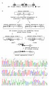
Pyramiding of the modifications/constructs created by the PCR-fusion/Gateway procedure. An example for pyramiding of gene modifications engineered by the PCR-fusion/Gateway procedure Showing the construction of an AtCesA7 cDNA mutant in which all eight Cys codons are mutated to Ser. Note that the pyramiding scheme described could be applied for all described cloning protocols. (A) General Cys residue structure of the Zn-finger domain of the CESA proteins. The two pairs of Cys residues which are mutated separately are designated. (B) Schematic of pyramiding of the Cys-residue mutagenesis in the Zn-finger domain of AtCesA7. Step 1: two AtCesA7Cys>Ser cDNA mutants are constructed in parallel, starting from wt AtCesA7 cDNA using the 'site-directed mutagenesis' protocol (Fig. 4a). In mutant expression clone 1 the codons of first pair of Cys are converted to Ser and in clone 2 the last pair. The mutated cDNA from expression clones 1 and 2 are generated using BP clonase and Gateway donor vector (pDONR/Zeo) to obtain mutant entry clones 1 and 2. Terminal regions from each of the mutated entry clones are PCR amplified with specific primers containing mutated codons of the remaining internal Cys residues of the Zn-finger. Overlap extension and cloning of the assembled DNA fragment into p3KC binary destination vector resulted in expression clone 3 in which all eight Cys codons are mutated to Ser. (C) Sequence analysis of the region of the Zn-finger domain mutant in expression clone 3. The converted Cys>Ser residues are marked with (*) and replaced nucleotides are shown in brackets.

References

    1. Hilson P. Cloned sequence repertoires for small- and large-scale biology. Trends Plant Sci. 2006;11:133–141. doi: 10.1016/j.tplants.2006.01.006. - DOI - PubMed
    1. Dafny-Yelin M, Tzfira T. Delivery of multiple transgenes to plant cells. Plant Physiol. 2007;145:1118–1128. doi: 10.1104/pp.107.106104. - DOI - PMC - PubMed
    1. Marsischky G, LaBaer J. Many paths to many clones: a comparative look at high-throughput cloning methods. Genome Res. 2004;14:2020–2028. doi: 10.1101/gr.2528804. - DOI - PubMed
    1. Lu Q. Seamless cloning and gene fusion. Trends Biotechnol. 2005;23:199–207. doi: 10.1016/j.tibtech.2005.02.008. - DOI - PMC - PubMed
    1. Hartley JL, Temple GF, Brasch MA. DNA cloning using in vitro site-specific recombination. Genome Res. 2000;10:1788–1795. doi: 10.1101/gr.143000. - DOI - PMC - PubMed

LinkOut - more resources