

The development of 3-D, in vitro, endothelial culture models for the study of coronary artery disease
- PMID: 19863806
- PMCID: PMC2773771
- DOI: 10.1186/1475-925X-8-30
The development of 3-D, in vitro, endothelial culture models for the study of coronary artery disease
Abstract
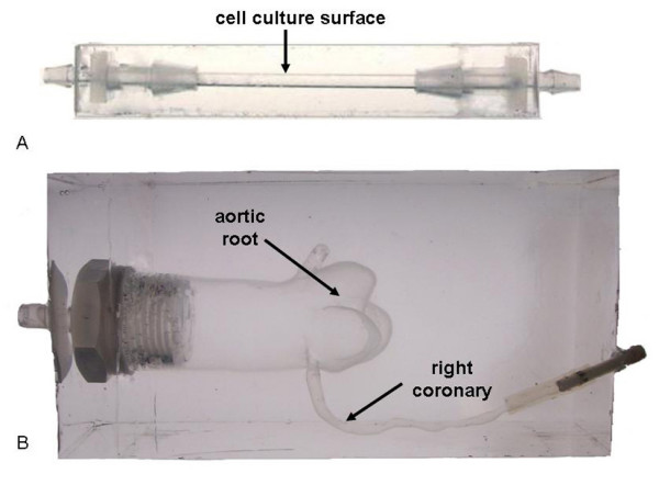
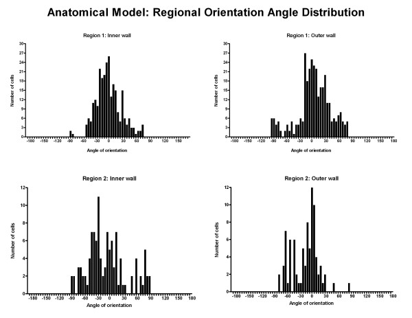
The response of the vascular endothelium to wall shear stress plays a central role in the development and progression of atherosclerosis. Current studies have investigated endothelial response using idealized in vitro flow chambers. Such cell culture models are unable to accurately replicate the complex in vivo wall shear stress patterns arising from anatomical geometries. To better understand this implication, we have created both simplified/tubular and anatomically realistic in vitro endothelial flow models of the human right coronary artery. A post-mortem vascular cast of the human left ventricular outflow tract was used to create geometrically accurate silicone elastomer models. Straight, tubular models were created using a custom made mold. Following the culture of human abdominal aortic endothelial cells within the inner lumen, cells were exposed to steady flow (Re = 233) for varying time periods. The resulting cell morphology was analyzed in terms of shape index and angle of orientation relative to the flow direction. In both models a progressive elongation and alignment of the endothelium in the flow direction was observed following 8, 12, and 24 hours. This change, however, was significantly less pronounced in the anatomical model (as observed from morphological variations indicative of localized flow features). Differences were also observed between the inner and outer walls at the disease-prone proximal region. Since morphological adaptation is a visual indication of endothelial shear stress activation, the use of anatomical models in endothelial genetic and biochemical studies may offer better insight into the disease process.
Figures


Similar articles
-
Endothelial cell morphologic response to asymmetric stenosis hemodynamics: effects of spatial wall shear stress gradients.J Biomech Eng. 2010 Aug;132(8):081013. doi: 10.1115/1.4001891. J Biomech Eng. 2010. PMID: 20670062
-
Coronary arteries hemodynamics: effect of arterial geometry on hemodynamic parameters causing atherosclerosis.Med Biol Eng Comput. 2020 Aug;58(8):1831-1843. doi: 10.1007/s11517-020-02185-x. Epub 2020 Jun 9. Med Biol Eng Comput. 2020. PMID: 32519006
-
Vascular cell adhesion molecule-1 expression in endothelial cells exposed to physiological coronary wall shear stresses.J Biomech Eng. 2009 Aug;131(8):081003. doi: 10.1115/1.3148191. J Biomech Eng. 2009. PMID: 19604015
-
Prediction of sites of coronary atherosclerosis progression: In vivo profiling of endothelial shear stress, lumen, and outer vessel wall characteristics to predict vascular behavior.Curr Opin Cardiol. 2003 Nov;18(6):458-70. doi: 10.1097/00001573-200311000-00007. Curr Opin Cardiol. 2003. PMID: 14597887 Review.
-
Microfluidic models of vascular functions.Annu Rev Biomed Eng. 2012;14:205-30. doi: 10.1146/annurev-bioeng-071811-150052. Epub 2012 Apr 23. Annu Rev Biomed Eng. 2012. PMID: 22540941 Review.
Cited by
-
Emerging trends in multiscale modeling of vascular pathophysiology: Organ-on-a-chip and 3D printing.Biomaterials. 2019 Mar;196:2-17. doi: 10.1016/j.biomaterials.2018.07.029. Epub 2018 Jul 23. Biomaterials. 2019. PMID: 30072038 Free PMC article. Review.
-
Increased MMP activity in curved geometries disrupts the endothelial cell glycocalyx creating a proinflammatory environment.PLoS One. 2018 Aug 23;13(8):e0202526. doi: 10.1371/journal.pone.0202526. eCollection 2018. PLoS One. 2018. PMID: 30138400 Free PMC article.
-
Soluble CD54 induces human endothelial cells ex vivo expansion useful for cardiovascular regeneration and tissue engineering application.Int J Cardiol Heart Vasc. 2015 Jan 9;6:48-53. doi: 10.1016/j.ijcha.2015.01.004. eCollection 2015 Mar 1. Int J Cardiol Heart Vasc. 2015. PMID: 28785626 Free PMC article.
-
Glycocalyx Degradation Induces a Proinflammatory Phenotype and Increased Leukocyte Adhesion in Cultured Endothelial Cells under Flow.PLoS One. 2016 Dec 1;11(12):e0167576. doi: 10.1371/journal.pone.0167576. eCollection 2016. PLoS One. 2016. PMID: 27907146 Free PMC article.
-
Effect of manufacturing and experimental conditions on the mechanical and surface properties of silicone elastomer scaffolds used in endothelial mechanobiological studies.Biomed Eng Online. 2017 Jul 14;16(1):90. doi: 10.1186/s12938-017-0380-5. Biomed Eng Online. 2017. PMID: 28705250 Free PMC article.
References
-
- Rosamond W, Flegal K, Friday G, Furie K, Go A, Greenlund K, Haase N, Ho M, Howard V, Kissela B, et al. Heart Disease and Stroke Statistics--2007 Update: A Report From the American Heart Association Statistics Committee and Stroke Statistics Subcommittee. Circulation. 2007;115:e69–171. doi: 10.1161/CIRCULATIONAHA.106.179918. - DOI - PubMed
Publication types
MeSH terms
Substances
LinkOut - more resources
Full Text Sources
Medical
Miscellaneous
