Induction of sperm head abnormalities by incorporated radionuclides: dependence on subcellular distribution, type of radiation, dose rate, and presence of radioprotectors
- PMID: 1986404
- PMCID: PMC5397899
Induction of sperm head abnormalities by incorporated radionuclides: dependence on subcellular distribution, type of radiation, dose rate, and presence of radioprotectors
Abstract
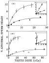
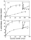
In contrast to the biological effects caused by exposure to external beams of radiation, the effects of tissue-incorporated radionuclides are highly dependent on the type of radiation emitted and on their distribution at the macroscopic, microscopic, and subcellular levels, which are in turn determined by the chemical nature of the radionuclides administered. Induction of abnormalities of sperm heads in mice is investigated in this work after the injection of a variety of radiochemicals including alpha emitters. When the initial slopes of the dose-response curves are used to compare the relative biological effectiveness (RBE) of different radiocompounds, the alpha particles emitted in the decay of 210Po are more effective than Auger electrons emitted by 125I incorporated in the DNA of the spermatogonial cells, and both emissions are more effective than X rays. It is also shown that the Auger emitters (125I, 111In) distributed in the cell nucleus are more efficient in producing abnormalities than the same radionuclides localized in the cytoplasm. These findings are consistent with our earlier observations, where spermatogonial cell survival is assayed as a function of the testicular absorbed dose. Further, chronic irradiation of testis with gamma rays from intratesticularly administered 7Be is about three times more effective in causing abnormalities than a single acute exposure to 120-kVp X rays. The resulting RBE values correlate well with our data on sperm head survival with the same radiocompounds. Finally, the radioprotector cysteamine, when administered in small, nontoxic amounts, significantly reduces the incidence of sperm abnormalities from alpha-particle radiation as well as emissions from 125I incorporated into DNA, the dose reduction factors being 10 and 14, respectively.
Figures


Similar articles
-
Biological consequence of nuclear versus cytoplasmic decays of 125I: cysteamine as a radioprotector against Auger cascades in vivo.Radiat Res. 1990 Nov;124(2):188-93. Radiat Res. 1990. PMID: 2247599
-
Relative biological effectiveness of alpha-particle emitters in vivo at low doses.Radiat Res. 1994 Mar;137(3):352-60. Radiat Res. 1994. PMID: 8146279 Free PMC article.
-
Radiotoxicity of some iodine-123, iodine-125 and iodine-131-labeled compounds in mouse testes: implications for radiopharmaceutical design.J Nucl Med. 1992 Dec;33(12):2196-201. J Nucl Med. 1992. PMID: 1460515
-
The Auger electron effect in radiation dosimetry.Health Phys. 1994 Nov;67(5):471-6. doi: 10.1097/00004032-199411000-00002. Health Phys. 1994. PMID: 7928358 Review.
-
Dosimetry of Auger-electron-emitting radionuclides: report no. 3 of AAPM Nuclear Medicine Task Group No. 6.Med Phys. 1994 Dec;21(12):1901-15. doi: 10.1118/1.597227. Med Phys. 1994. PMID: 7700197 Review.
Cited by
-
Protection by DMSO against cell death caused by intracellularly localized iodine-125, iodine-131 and polonium-210.Radiat Res. 2000 Apr;153(4):416-27. doi: 10.1667/0033-7587(2000)153[0416:pbdacd]2.0.co;2. Radiat Res. 2000. PMID: 10761002 Free PMC article.
-
Biological effect of lead-212 localized in the nucleus of mammalian cells: role of recoil energy in the radiotoxicity of internal alpha-particle emitters.Radiat Res. 1994 Nov;140(2):276-83. Radiat Res. 1994. PMID: 7938477 Free PMC article.
-
On the equivalent dose for Auger electron emitters.Radiat Res. 1993 Apr;134(1):71-8. Radiat Res. 1993. PMID: 8475256 Free PMC article.
-
Design and testing of a microcontroller that enables alpha particle irradiators to deliver complex dose rate patterns.Phys Med Biol. 2018 Dec 18;63(24):245022. doi: 10.1088/1361-6560/aaf269. Phys Med Biol. 2018. PMID: 30524061 Free PMC article.
-
Quantitative γ-H2AX immunofluorescence method for DNA double-strand break analysis in testis and liver after intravenous administration of 111InCl3.EJNMMI Res. 2020 Mar 19;10(1):22. doi: 10.1186/s13550-020-0604-8. EJNMMI Res. 2020. PMID: 32189079 Free PMC article.
References
-
- Oakberg EF. Duration of spermatogenesis in the mouse and timing of stages of the cycle of the seminiferous epithelium. Am J Anat. 1956;99:507–516. - PubMed
-
- Oakberg EF. Sensitivity and time of degeneration of spermatogenic cells irradiated in various stages of maturation in the mouse. Radiat Res. 1955;2:369–391. - PubMed
-
- Meistrich ML. Critical components of testicular function and sensitivity to disruption. Biol Reprod. 1986;34:17–28. - PubMed
-
- Sastry KSR, Rao DV. Dosimetry of low energy electrons. In: Rao DV, Chandra R, Graham M, editors. Physics of Nuclear Medicine: Recent Advances. American Institute of Physics; New York: 1984. pp. 169–208. Medical Physics Monograph No. 10.
-
- Rao DV, Sastry KSR, Grimmond HE, Howell RW, Govelitz GF, Lanka VK, Mylavarapu VB. Cytotoxicity of some indium radiopharmaceuticals in mouse testes. J Nucl Med. 1988;29:375–384. - PubMed
Publication types
MeSH terms
Substances
Grants and funding
LinkOut - more resources
Full Text Sources
