. 2010 Feb;298(2):F314-22.
doi: 10.1152/ajprenal.00483.2009. Epub 2009 Oct 28.

Reductions in laminin beta2 mRNA translation are responsible for impaired IGFBP-5-mediated mesangial cell migration in the presence of high glucose

Affiliations

Reductions in laminin beta2 mRNA translation are responsible for impaired IGFBP-5-mediated mesangial cell migration in the presence of high glucose

Valerie Schaeffer et al. Am J Physiol Renal Physiol. 2010 Feb.

Abstract

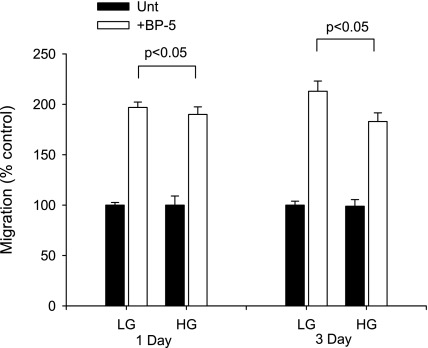
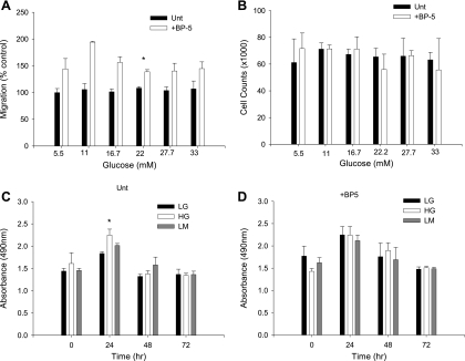
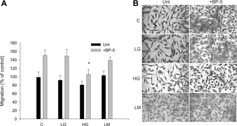
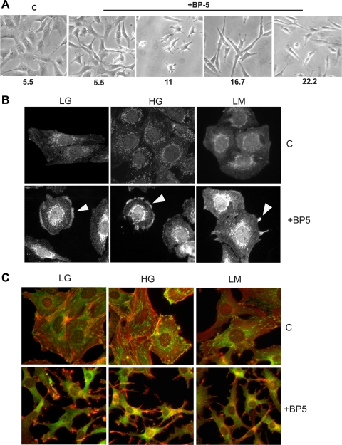
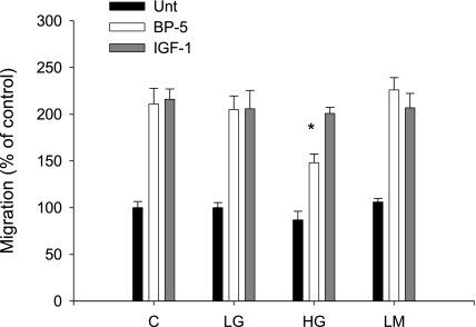
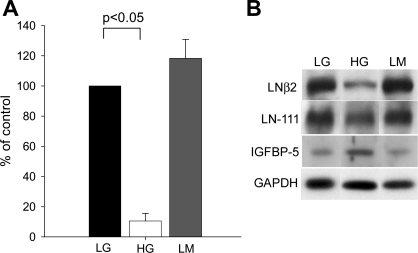
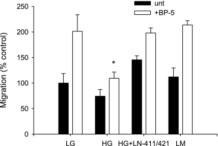
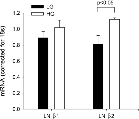
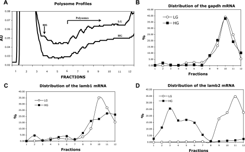
Insulin-like growth factor binding protein-5 (IGFBP-5) mediates mesangial cell migration through activation of cdc42, and laminin421 binding to alpha(6)beta(1)-integrin (Berfield AK, Hansen KM, Abrass CK. Am J Physiol Cell Physiol 291: C589-C599, 2006). Because glomerular expression of laminin beta(2) is reduced in diabetic rats (Abrass CK, Spicer D, Berfield AK, St. John PL, Abrahamson DR. Am J Pathol 151: 1131-1140, 1997), we directly examined the effect of hyperglycemia on mesangial cell migration and laminin beta2 expression. Migration mediated by IGFBP-5 is impaired in the presence of 25 mM glucose. This reduction in migration was found to result from a loss in mesangial cell synthesis of laminin421, and IGFBP-5-induced migration could be restored by replacing laminin421. Additional studies showed that there was selective reduction in mRNA translation of laminin beta2 in the presence of high glucose. Preserved synthesis of laminin beta1 indicates that not all proteins are reduced by high glucose and confirms prior data showing that laminin411 cannot substitute for laminin421 in IGFBP-5-mediated migration. Given the importance of mesangial migration in the reparative response to diabetes-associated mesangiolysis, these findings provide new insights into abnormalities associated with diabetic nephropathy and the potential importance of differential control of protein translation in determination of alterations of protein expression.


Figures

Fig. 1.
Fig. 1.
Effect of acute and chronic exposure to high glucose (HG). Mesangial cells (MC) were exposed to HG for 1 or 3 days before testing migratory response to insulin-like growth factor binding protein-5 (IGFBP-5). Differences in migratory response were only observed after 3 days of exposure to HG. LG, low glucose. *P < 0.05, ANOVA.
Fig. 2.
Fig. 2.
Glucose dose-response experiment. Effect is shown of 3 days of increasing glucose concentration on cell migration (A), cell numbers (B), and cell metabolism with (D) and without IGFBP-5 (C). LM, low-glucose+mannitol. *P < 0.05, ANOVA.
Fig. 3.
Fig. 3.
MC migration after 7 days in HG medium. MC were cultured for 7 days in control (C; 200 mg/dl glucose), 5.5 mM glucose (LG), 25 mM glucose (HG), or 5.5 mM glucose+mannitol (LM) medium before assessment of migration with and without IGFBP-5201-218 (30 μg/ml). A: migration was expressed as a % of control. *P < 0.05, ANOVA. B: photomicrograph showing cells filling the wounded area.
Fig. 4.
Fig. 4.
Cell phenotype, cdc42 activation, and cytoskeletal reorganization. A: MC were cultured in various concentrations of glucose for 3 days before addition of IGFBP-5201-218 and were photographed with phase-contrast microscopy 48 h later. In keeping with previous studies, the change in phenotype was unaffected by glucose concentration. B: MC were cultured for 7 days in 5.5 mM glucose (LG), 25 mM glucose (HG), or 5.5 mM glucose+mannitol (LM) medium before addition of IGFBP-5201-218 (30 μg/ml). One hour later, cells were stained with antibody to cdc42GAP. The activated cdc42-cdc42GAP complex becomes clustered and is detected in areas where the membrane is beginning to reorganize (arrowheads). C: 24 h after the addition of IGFBP-5201-218, F-actin (red) and vinculin (green) staining were performed. In untreated cultures in all 3 media, cells are spread and vinculin is stained in a submembrane array. F-actin fibers span the cell in both parallel and overlapping patterns. Following the addition of IGFBP-5201-218, cells have developed a spider-like morphology with long filopodia. The submembrane array of vinculin is lost, and F-actin has reorganized and extends along the filopodial projections. These responses to IGFBP-5 were not altered by changes in glucose concentration in the medium.
Fig. 5.
Fig. 5.
MC migration in response to IGFBP-5201-218 and IGF-1. MC migration was determined in control (C; 200 mg/dl glucose), 5.5 mM glucose (LG), 25 mM glucose (HG), or 5.5 mM glucose+mannitol (LM) with and without IGFBP-5201-218 (30 μg/ml) or IGF-1 (100 nM). MC were maintained in the experimental medium for 3 days before the migration assay. Bar graphs show results expressed as a % of control. *P < 0.05, ANOVA.
Fig. 6.
Fig. 6.
Laminin (LN) β2 protein. A: Western blot of proteins extracted from cells exposed to LG, HG, or LM for 10 days. Densitometric analysis of Western blots for laminin β2 was corrected for loading controls. Experiments were repeated on 4 separate occasions. *P < 0.05, ANOVA. B: representative Western blot for laminin β2, showing comparison with staining for laminin β1/γ1 as detected by antibodies to LN111, IGFBP-5, and GAPDH.
Fig. 7.
Fig. 7.
Restoration of IGFBP-5-induced MC migration by plating on LN421. MC were exposed to varying concentrations of glucose for 10 days followed by assessment of migration in response to IGFBP-5201-218 after plating on the substrates noted. The reduction in MC migration in HG was restored when MC were plated on matrix containing LN421 in addition to LN411. *P < 0.05, ANOVA.
Fig. 8.
Fig. 8.
Quantification of laminin β1 and laminin β2 mRNA. MC were exposed to LG or HG for 10 days. LNβ1 and LNβ2 mRNA content were determined by quantitative real-time PCR. mRNA content corrected for 18S is shown; n = 3/condition, each run in triplicate. *P < 0.05, ANOVA.
Fig. 9.
Fig. 9.
mRNA association with polysomes. MC were cultured in LG or HG for 7 days before extraction. Polysomes were fractionated on a sucrose gradient, and each fraction was assayed for specific mRNA content by qPCR. In A, a slight reduction is shown in the total amount of polysomes in the high-molecular-weight fractions in HG-treated MC compared with LG, which is consistent with a reduction in total protein synthesis. Monosomes are at the left, and polysomes of increasing size are to the right. The profiles for gapdh (B), lamb1 (C), and lamb2 (D) are shown. Note that for both gapdh and lamb1, curves are similar in both LG and HG. In contrast, lamb2 mRNA is not detected in high-molecular-weight polysome fractions in HG, indicating that translation of this mRNA is impaired by HG.

References

    1. Abrass CK, Berfield AK, Andress DL. Heparin binding domain of insulin-like growth factor binding protein-5 stimulates mesangial cell migration. Am J Physiol Renal Physiol 273: F899–F906, 1997 - PubMed
    1. Abrass CK, Spicer D, Berfield AK, St. John PL, Abrahamson DR. Diabetes induces changes in glomerular development and laminin b2 (s-laminin) expression. Am J Pathol 151: 1131–1140, 1997 - PMC - PubMed
    1. Abrass CK, Spicer D, Raugi GJ. Insulin induces a change in extracellular matrix glycoproteins synthesized by rat mesangial cells in culture. Kidney Int 46: 613–620, 1994 - PubMed
    1. Abrass CK, Spicer D, Raugi GJ. Induction of nodular sclerosis by insulin in rat mesangial cells in vitro: studies of collagen. Kidney Int 47: 25–37, 1995 - PubMed
    1. Bell L, Madri JA. Influence of the angiotensin system on endothelial and smooth muscle cell migration. Am J Pathol 137: 7–12, 1990 - PMC - PubMed

Publication types

MeSH terms