. 2010 Jan;298(1):F177-86.
doi: 10.1152/ajprenal.00464.2009. Epub 2009 Oct 28.

Angiotensin II stimulates trafficking of NHE3, NaPi2, and associated proteins into the proximal tubule microvilli

Affiliations

Angiotensin II stimulates trafficking of NHE3, NaPi2, and associated proteins into the proximal tubule microvilli

Anne D M Riquier-Brison et al. Am J Physiol Renal Physiol. 2010 Jan.

Abstract

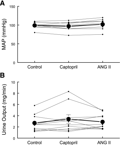
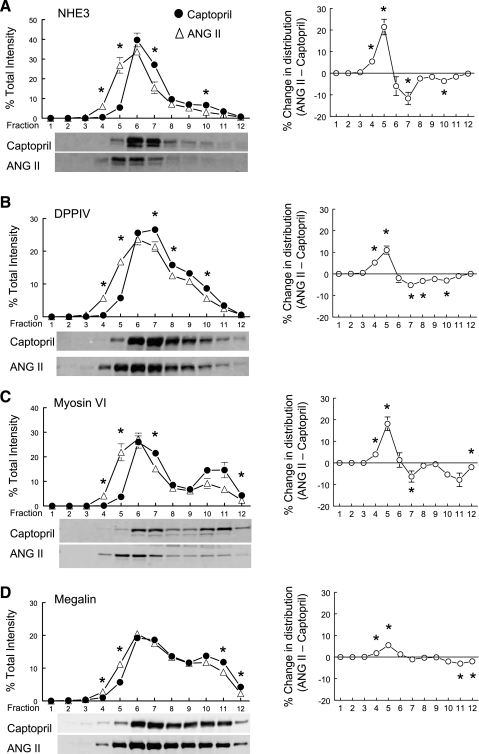
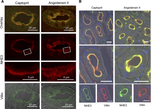
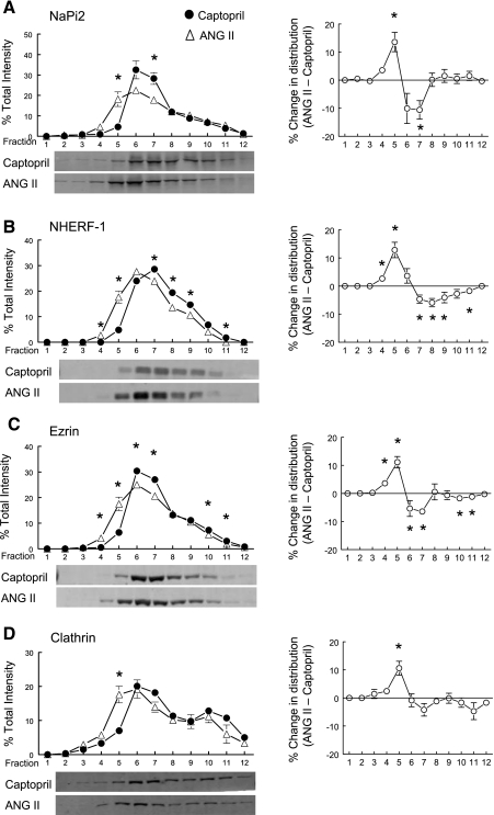
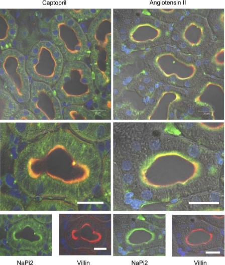
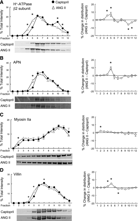
Angiotensin II (ANG II) stimulates proximal tubule (PT) sodium and water reabsorption. We showed that treating rats acutely with the angiotensin-converting enzyme inhibitor captopril decreases PT salt and water reabsorption and provokes rapid redistribution of the Na(+)/H(+) exchanger isoform 3 (NHE3), Na(+)/Pi cotransporter 2 (NaPi2), and associated proteins out of the microvilli. The aim of the present study was to determine whether acute ANG II infusion increases the abundance of PT NHE3, NaPi2, and associated proteins in the microvilli available for reabsorbing NaCl. Male Sprague-Dawley rats were infused with a dose of captopril (12 microg/min for 20 min) that increased PT flow rate approximately 20% with no change in blood pressure (BP) or glomerular filtration rate (GFR). When ANG II (20 ng x kg(-1) x min(-1) for 20 min) was added to the captopril infusate, PT volume flow rate returned to baseline without changing BP or GFR. After captopril, NHE3 was localized to the base of the microvilli and NaPi2 to subapical cytoplasmic vesicles; after 20 min ANG II, both NHE3 and NaPi2 redistributed into the microvilli, assayed by confocal microscopy and density gradient fractionation. Additional PT proteins that redistributed into low-density microvilli-enriched membranes in response to ANG II included myosin VI, DPPIV, NHERF-1, ezrin, megalin, vacuolar H(+)-ATPase, aminopeptidase N, and clathrin. In summary, in response to 20 min ANG II in the absence of a change in BP or GFR, multiple proteins traffic into the PT brush-border microvilli where they likely contribute to the rapid increase in PT salt and water reabsorption.


Figures

Fig. 1.
Fig. 1.
Effects of captopril and ANG II infusion protocols on mean arterial pressure (MAP) and urine output. Rats were sequentially infused with 4.0% BSA in 0.9% saline (50 μl/min) for >30 min, then with captopril (12 μg/min) for 20 min, and then with ANG II + captopril (20 ng·kg−1·min−1 ANG II and 12 μg/min captopril). A: MAP was measured continuously at the carotid (mmHg). B: urine was collected continuously in 10-min intervals and output rate was averaged over the treatment period (mg/min), n = 10, individual traces and means ± SE.
Fig. 2.
Fig. 2.
Effects of acute ANG II infusion on the density distribution of proximal tubule microvillar proteins: Na+/H+ exchanger isoform 3 (NHE3; A), dipeptidyl peptidase IV (DPPIV; B), myosin VI (C), and megalin (D). Comparison of protein density distribution between 20-min captopril infusion (12 μg/min; ·, n = 5) and 20-min ANG II (20 ng·kg−1·min−1 ANG II and 12 μg/min captopril; ▵). Immunoreactivity in each fraction is expressed as a percentage of total signal in all 12 fractions. AD, right: summarize the difference in the density distribution between ANG II- and captopril-treated groups. Values are means ± SE. *P < 0.05 compared with corresponding captopril fraction, unpaired Student's t-test after ANOVA. Shown below AD are representative immunoblots of corresponding protein from typical experiments with NHE3 detected at 83 kDa, DPPIV at 89 kDa, myosin VI at 145 kDa, and megalin at 345 kDa.
Fig. 3.
Fig. 3.
Indirect immunofluorescence microscopy of NHE3 redistribution in the proximal tubule was conducted in rats infused acutely with captopril (left) or ANG II (right) in distinct series conducted without controlling arterial pressure (A) or with controlling arterial pressure (B) as described in experimental procedures. In both series, a kidney sample from each group was placed on the same slide and processed identically for detection of NHE3 and villin. A: anti-NHE3 was detected with AlexaFluor 568-conjugated goat anti-rabbit secondary antibody and anti-villin with AlexaFluor 488-conjugated goat anti-mouse secondary antibody. B: anti-NHE3 was detected with FITC-conjugated goat anti-rabbit secondary antibody and anti-villin with Alexa 568-conjugated goat anti-mouse secondary and bar is 20 μm.
Fig. 4.
Fig. 4.
Effects of acute ANG II infusion on the density distribution of proximal tubule microvillar proteins: NaPi2 (A), NHE3 regulatory factor 1 (NHERF-1; B), ezrin (C), and clathrin (D). Comparison of protein density distribution between 20-min captopril infusion (12 μg/min; ·, n = 5) and 20-min ANG II (20 ng·kg−1·min−1 ANG II and 12 μg/min captopril; ▵). Immunoreactivity in each fraction is expressed as a percentage of total signal in all 12 fractions. AD, right: summarize the difference in the density distribution between ANG II- and captopril-treated groups. Values are means ± SE. *P < 0.05 compared with corresponding captopril fraction, unpaired Student's t-test after ANOVA. Shown below AD are representative immunoblots of corresponding protein from typical experiments with NaPi2 detected at 82 kDa, NHERF-1 at 50 kDa, ezrin at 69 kDa, and clathrin at 193 kDa.
Fig. 5.
Fig. 5.
Indirect immunofluorescence microscopy of Na+/Pi cotransporter 2 (NaPi2) redistribution in the proximal tubule of rats infused acutely with captopril (left) or ANG II (right) and perfusion fixed without elevating arterial pressure as described in experimental procedures. A kidney sample from each group was placed on the same slide and processed identically for detection of NaPi2 and villin. Anti-NaPi2 was detected with FITC-conjugated goat anti-rabbit secondary antibody and anti-villin with Alexa 568-conjugated goat anti-mouse secondary. Bar is 20 μm.
Fig. 6.
Fig. 6.
Effects of acute ANG II infusion on the density distribution of proximal tubule microvillar proteins: vacuolar H+-ATPase β2 (A), aminopeptidase N (APN; B), myosin IIa (C), and villin (D). Comparison of protein density distribution between 20-min captopril infusion (12 μg/min; ·, n = 5) and 20-min ANG II (20 ng·kg−1·min−1 ANG II and 12 μg/min captopril; ▵). Immunoreactivity in each fraction is expressed as a percentage of total signal in all 12 fractions. AD, right: summarize the difference in the density distribution between ANG II- and captopril-treated groups. Values are means ± SE. *P < 0.05 compared with corresponding captopril fraction, unpaired Student's t-test after ANOVA. Shown below AD are representative immunoblots of corresponding protein from typical experiments with vacuolar H+-ATPase β2 subunit detected at 57 kDa, APN at 110 kDa, myosin IIa detected at 227 kDa, and villin detected at 95 kDa.

References

    1. Ardaillou R. Active fragments of angiotensin II: enzymatic pathways of synthesis and biological effects. Curr Opin Nephrol Hypertens 6: 28–34, 1997 - PubMed
    1. Arrondel C, Vodovar N, Knebelmann B, Grunfeld JP, Gubler MC, Antignac C, Heidet L. Expression of the nonmuscle myosin heavy chain IIA in the human kidney and screening for MYH9 mutations in Epstein and Fechtner syndromes. J Am Soc Nephrol 13: 65–74, 2002 - PubMed
    1. Biemesderfer D, Nagy T, DeGray B, Aronson PS. Specific association of megalin and the Na+/H+ exchanger isoform NHE3 in the proximal tubule. J Biol Chem 274: 17518–17524, 1999 - PubMed
    1. Braam B, Allen P, Benes E, Koomans HA, Navar LG, Hammond T. Human proximal tubular cell responses to angiotensin II analyzed using DNA microarray. Eur J Pharmacol 464: 87–94, 2003 - PubMed
    1. Brown D, Hirsch S, Gluck S. Localization of a proton-pumping ATPase in rat kidney. J Clin Invest 82: 2114–2126, 1988 - PMC - PubMed

Publication types

MeSH terms