. 2010 Apr;82(4):791-801.
doi: 10.1095/biolreprod.109.081059. Epub 2009 Oct 28.

Genomic profiling of microRNAs and messenger RNAs reveals hormonal regulation in microRNA expression in human endometrium

Affiliations

Genomic profiling of microRNAs and messenger RNAs reveals hormonal regulation in microRNA expression in human endometrium

Satu Kuokkanen et al. Biol Reprod. 2010 Apr.

Abstract

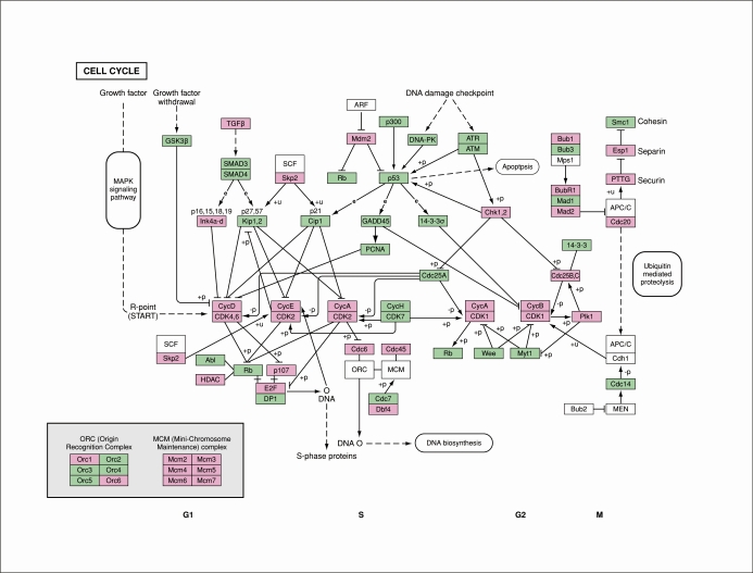
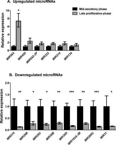
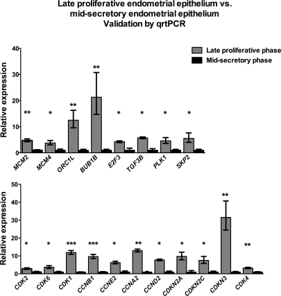
MicroRNAs (miRNAs), a class of small noncoding RNAs that regulate gene expression, have fundamental roles in biological processes, including cell differentiation and proliferation. These small molecules mainly direct either target messenger RNA (mRNA) degradation or translational repression, thereby functioning as gene silencers. Epithelial cells of the uterine lumen and glands undergo cyclic changes under the influence of the sex steroid hormones estradiol-17beta and progesterone. Because the expression of miRNAs in human endometrium has been established, it is important to understand whether miRNAs have a physiological role in modulating the expression of hormonally induced genes. The studies herein establish concomitant differential miRNA and mRNA expression profiles of uterine epithelial cells purified from endometrial biopsy specimens in the late proliferative and midsecretory phases. Bioinformatics analysis of differentially expressed mRNAs revealed cell cycle regulation as the most significantly enriched pathway in the late proliferative-phase endometrial epithelium (P = 5.7 x 10(-15)). In addition, the WNT signaling pathway was enriched in the proliferative phase. The 12 miRNAs (MIR29B, MIR29C, MIR30B, MIR30D, MIR31, MIR193A-3P, MIR203, MIR204, MIR200C, MIR210, MIR582-5P, and MIR345) whose expression was significantly up-regulated in the midsecretory-phase samples were predicted to target many cell cycle genes. Consistent with the role of miRNAs in suppressing their target mRNA expression, the transcript abundance of predicted targets, including cyclins and cyclin-dependent kinases, as well as E2F3 (a known target of MIR210), was decreased. Thus, our findings suggest a role for miRNAs in down-regulating the expression of some cell cycle genes in the secretory-phase endometrial epithelium, thereby suppressing cell proliferation.


Figures

FIG. 1.
FIG. 1.
Dendrogram and unsupervised hierarchical clustering. Expression data from all mRNA probes (A) and expression data from differentially expressed miRNAs showing a greater than 1.5-fold or less than 1.5-fold change in expression with P < 0.05 (B) in four late proliferative-phase (pp) and four midsecretory-phase (sp) samples. The top dendrogram shows the relationship between the samples based on gene expression patterns. Hierarchical clustering analyses robustly separate the late proliferative-phase samples from the midsecretory-phase samples and assign each sample to the correct menstrual cycle phase. The expression intensity of each gene in each sample varies from high (red) to low (green).
FIG. 2.
FIG. 2.
Transcripts regulating the mammalian cell cycle are up-regulated in the proliferative-phase endometrial epithelium. Genes with a greater than 2-fold increase in transcript abundance with P < 0.05 in the late proliferative-phase samples compared with the midsecretory-phase samples from the human genome U133 Plus 2.0 arrays are indicated in purple boxes and represent 38 up-regulated genes. Green and white boxes indicate cell cycle genes that showed no differential expression between the late proliferative-phase samples compared with the midsecretory-phase samples. The diagram is based on the cell cycle pathway in the KEGG pathway database [34, 35].
FIG. 3.
FIG. 3.
Transcripts mediating cell cycle regulation are differentially regulated between the late proliferative-phase vs. midsecretory-phase endometrial epithelium. Differentially expressed transcripts were subjected to bioinformatics analysis using IPA 7.6. A) Most significantly enriched groups relating to molecular and cellular functions. B) Most significantly enriched canonical signaling pathways. The total number of genes in a pathway is boldfaced. For each pathway, the percentage of genes that are of decreased (green) or increased (red) transcript abundance is indicated at the top. Portions of pathway genes that are not significantly changed are also shown (white). The yellow square for each pathway indicates –log(P value). The yellow arrow indicates P = 0.05.
FIG. 4.
FIG. 4.
Expression of selected up-regulated (A) and down-regulated (B) miRNAs in the late proliferative-phase endometrial samples relative to the midsecretory-phase endometrial samples by quantitative real-time PCR. All samples were normalized to RNU48. Relative gene expression of the late proliferative-phase vs. midsecretory-phase samples was assessed using the 2−ΔΔCT method [33]. Data shown indicate relative expression of the late proliferative-phase samples (gray bar) with respect to the midsecretory-phase samples (black bars) set to 1. The error bars show the SEM. *P < 0.05, **P < 0.01, and ***P < 0.001.
FIG. 5.
FIG. 5.
Quantitative real-time PCR validates gene expression array data from the cell cycle regulation ontological group. Nineteen genes were validated as differentially regulated cell cycle members from the human genome U133 Plus 2.0 array using quantitative real-time PCR. All samples were normalized to the housekeeping gene GAPDH. Data shown indicate relative expression of the late proliferative-phase samples (gray bar) with respect to the midsecretory-phase samples (black bars) set to 1. The error bars show the SEM. ***P < 0.001, **P < 0.01, and *P < 0.05.

Comment in

References

    1. Kurita T, Medina R, Schabel AB, Young P, Gama P, Parekh TV, Brody J, Cunha GR, Osteen KG, Bruner-Tran KL, Gold LI.The activation function-1 domain of estrogen receptor alpha in uterine stromal cells is required for mouse but not human uterine epithelial response to estrogen. Differentiation 2005; 73: 313–322. - PubMed
    1. Zhu L, Pollard JW.Estradiol-17beta regulates mouse uterine epithelial cell proliferation through insulin-like growth factor 1 signaling. Proc Natl Acad Sci U S A 2007; 104: 15847–15851. - PMC - PubMed
    1. Guzeloglu Kayisli O, Kayisli UA, Luleci G, Arici A.In vivo and in vitro regulation of Akt activation in human endometrial cells is estrogen dependent. Biol Reprod 2004; 71: 714–721. - PubMed
    1. Tong W, Pollard JW.Progesterone inhibits estrogen-induced cyclin D1 and cdk4 nuclear translocation, cyclin E- and cyclin A-cdk2 kinase activation, and cell proliferation in uterine epithelial cells in mice. Mol Cell Biol 1999; 19: 2251–2264. - PMC - PubMed
    1. Chen B, Pan H, Zhu L, Deng Y, Pollard JW.Progesterone inhibits the estrogen-induced phosphoinositide 3-kinase–>AKT–>GSK-3beta–>cyclin D1–>pRB pathway to block uterine epithelial cell proliferation. Mol Endocrinol 2005; 19: 1978–1990. - PubMed

Publication types

MeSH terms