

Analysis of herpes simplex virus type 1 DNA packaging signal mutations in the context of the viral genome
- PMID: 19864384
- PMCID: PMC2798400
- DOI: 10.1128/JVI.01489-09
Analysis of herpes simplex virus type 1 DNA packaging signal mutations in the context of the viral genome
Abstract
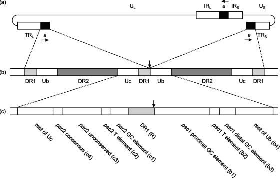
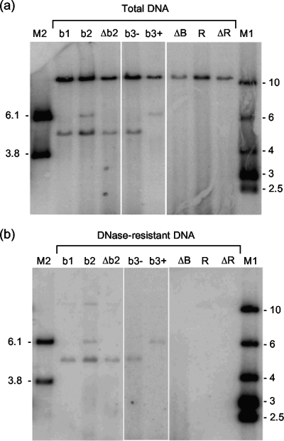
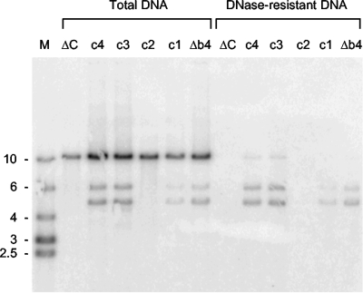
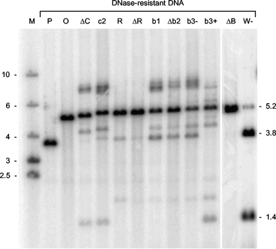
The minimal signal required for the cleavage and packaging of replicated concatemeric herpes simplex virus type 1 (HSV-1) DNA corresponds to an approximately 200-bp fragment, Uc-DR1-Ub, spanning the junction of the genomic L and S segments. Uc and Ub occupy positions adjacent to the L and S termini and contain motifs (pac2 and pac1, respectively) that are conserved near the ends of other herpesvirus genomes. We have used homologous Red/ET recombination in Escherichia coli to introduce wild-type and specifically mutated Uc-DR1-Ub fragments into an ectopic site of a cloned HSV-1 genome from which the resident packaging signals had been previously deleted. The resulting constructs were transfected into mammalian cells, and their abilities to replicate and become encapsidated, generate Uc- and Ub-containing terminal fragments, and give rise to progeny virus were assessed. In general, the results obtained agree well with previous observations made using amplicons and confirm roles for the pac2 T element in the initiation of DNA packaging and for the GC-rich motifs flanking the pac1 T element in termination. In contrast to a previous report, the sequence of the DR1 element was also crucial for DNA packaging. Following repair of the resident packaging signals in mammalian cells, recombination occurred at high frequency in progeny virus between the repaired sequences and mutated Uc-DR1-Ub inserts. This restored the ability of mutated Uc-DR1-Ub inserts to generate terminal fragments, although these were frequently larger than expected from simple repair of the original lesion.
Figures
Similar articles
-
Effects of mutations within the herpes simplex virus type 1 DNA encapsidation signal on packaging efficiency.J Virol. 2001 Oct;75(19):8977-86. doi: 10.1128/JVI.75.19.8977-8986.2001. J Virol. 2001. PMID: 11533161 Free PMC article.
-
Excision of DNA fragments corresponding to the unit-length a sequence of herpes simplex virus type 1 and terminus variation predominate on one side of the excised fragment.J Virol. 1994 Jul;68(7):4377-83. doi: 10.1128/JVI.68.7.4377-4383.1994. J Virol. 1994. PMID: 8207811 Free PMC article.
-
Herpes simplex virus type 1 DNA amplified as bacterial artificial chromosome in Escherichia coli: rescue of replication-competent virus progeny and packaging of amplicon vectors.Hum Gene Ther. 1998 Dec 10;9(18):2787-94. doi: 10.1089/hum.1998.9.18-2787. Hum Gene Ther. 1998. PMID: 9874276
-
Sequences within the herpesvirus-conserved pac1 and pac2 motifs are required for cleavage and packaging of the murine cytomegalovirus genome.J Virol. 1998 Jan;72(1):48-56. doi: 10.1128/JVI.72.1.48-56.1998. J Virol. 1998. PMID: 9420199 Free PMC article.
-
Herpes simplex virus type 1 replication and recombination.Biochimie. 1995;77(10):787-95. doi: 10.1016/0300-9084(96)88197-1. Biochimie. 1995. PMID: 8824776 Review.
Cited by
-
A Domain of Herpes Simplex Virus pUL33 Required To Release Monomeric Viral Genomes from Cleaved Concatemeric DNA.J Virol. 2017 Sep 27;91(20):e00854-17. doi: 10.1128/JVI.00854-17. Print 2017 Oct 15. J Virol. 2017. PMID: 28747509 Free PMC article.
-
A mutation in UL15 of herpes simplex virus 1 that reduces packaging of cleaved genomes.J Virol. 2011 Nov;85(22):11972-80. doi: 10.1128/JVI.00857-11. Epub 2011 Aug 31. J Virol. 2011. PMID: 21880766 Free PMC article.
-
Complete Genome Sequence of Herpes Simplex Virus 2 Strain G.Viruses. 2022 Mar 5;14(3):536. doi: 10.3390/v14030536. Viruses. 2022. PMID: 35336943 Free PMC article.
-
Herpesvirus BACs: past, present, and future.J Biomed Biotechnol. 2011;2011:124595. doi: 10.1155/2011/124595. Epub 2010 Oct 27. J Biomed Biotechnol. 2011. PMID: 21048927 Free PMC article. Review.
-
Cryo-EM structures of herpes simplex virus type 1 portal vertex and packaged genome.Nature. 2019 Jun;570(7760):257-261. doi: 10.1038/s41586-019-1248-6. Epub 2019 May 29. Nature. 2019. PMID: 31142842 Free PMC article.
References
-
- Baines, J. D., and S. K. Weller. 2005. Cleavage and packaging of herpes simplex virus 1 DNA, p. 135-150. In C. E. Catalano (ed.), Viral genome packaging machines: genetics, structure and mechanism. Kluwer Academic/Plenum Publishers, New York, NY.
-
- Broll, H., H.-J. Buhk, W. Zimmermann, and M. Goltz. 1999. Structure and function of the prDNA and genomic termini of the γ2-herpesvirus bovine herpesvirus type 4. J. Gen. Virol. 80:979-986. - PubMed
-
- Brown, J. C., M. A. McVoy, and F. L. Homa. 2002. Packaging DNA into herpesvirus capsids, p. 111-153. In A. Holzenburg and E. Bogner (ed.), Structure-function relationships of human pathogenic viruses. Kluwer Academic/Plenum Publishers, New York, NY.
-
- Chou, J., and B. Roizman. 1985. Isomerization of herpes simplex virus 1 genome: identification of the cis-acting and recombination sites within the domain of the a sequence. Cell 41:803-811. - PubMed
MeSH terms
Substances
Grants and funding
LinkOut - more resources
Full Text Sources
Other Literature Sources
Miscellaneous
