. 2010 Jan;84(1):387-96.
doi: 10.1128/JVI.01921-09.

Production and function of the cytoplasmic deproteinized relaxed circular DNA of hepadnaviruses

Affiliations

Production and function of the cytoplasmic deproteinized relaxed circular DNA of hepadnaviruses

Haitao Guo et al. J Virol. 2010 Jan.

Abstract

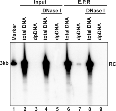
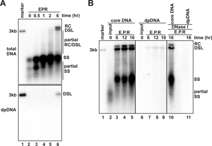
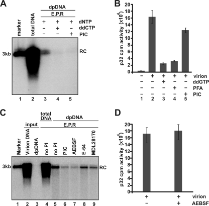
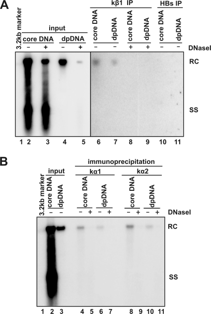
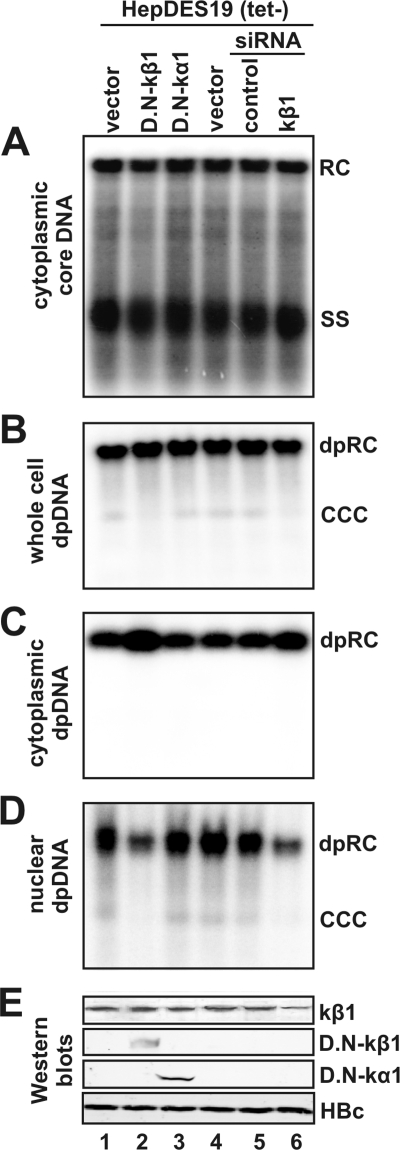
Removal of genome-bound viral DNA polymerase ought to be an essential step in the formation of hepadnavirus covalently closed circular DNA (cccDNA). We previously demonstrated that deproteinized (DP) relaxed circular DNA (rcDNA) of hepatitis B virus (HBV) existed in both the cytoplasm and nuclei of infected cells and the vast majority of cytoplasmic DP rcDNA was associated with DNase I-permeable nucleocapsids. In our efforts to investigate the role of the cytoplasmic DP rcDNA in cccDNA formation, we demonstrated that rcDNA deproteinization could occur in an endogenous DNA polymerase reaction with either virion-derived or intracellular nucleocapsids. As observed in the cytoplasm of virally infected cells, in vitro deproteinization requires the maturation of plus-strand DNA and results in changes in nucleocapsid structure that render the DP rcDNA susceptible to DNase I digestion. Remarkably, we found that the cytoplasmic DP rcDNA-containing nucleocapsids could be selectively immunoprecipitated with an antibody against the carboxyl-terminal peptide of HBV core protein and are associated with cellular nuclear transport receptors karyopherin-alpha and -beta. Moreover, transfection of small interfering RNA targeting karyopherin-beta1 mRNA or expression of a dominant-negative karyopherin-beta1 in a stable cell line supporting HBV replication resulted in the accumulation of DP rcDNA in cytoplasm and reduction of nuclear DP rcDNA and cccDNA. Our results thus favor a hypothesis that completion of plus-strand DNA synthesis triggers the genomic DNA deproteinization and structural changes of nucleocapsids, which leads to the exposure of nuclear localization signals in the C terminus of core protein and mediates the nuclear transportation of DP rcDNA via interaction with karyopherin-alpha and -beta.


Figures

FIG. 1.
FIG. 1.
Removal of genome-bounded polymerase and capsid disassembly occur following endogenous polymerase reaction in vitro. Approximately 108 DHBV virion particles prepared from DHBV-positive duck serum were contained in each 100-μl EPR mixture including 150 mM NaCl, 50 mM Tris-HCl (pH 8.0), 10 mM MgCl2, 1 mM DTT, 0.1% NP-40, and 0.1 mM dNTP. The reaction mixtures were either without incubation (lanes 2 to 5) or incubated at 37°C for 16 h (lanes 6 to 9), followed by total DNA and DP DNA extraction without (lanes 2, 3, 6, and 7) or with (lanes 4, 5, 8 and 9) prior DNase I digestion, respectively. The viral DNA were resolved in agarose gel and detected by Southern blot hybridization with full-length riboprobe recognizing minus-strand DNA. One hundred picograms of 3.0-kb unit-length DHBV DNA served as the quantification standard and molecular weight marker (lane 1). The position of rcDNA (RC) is indicated.
FIG. 2.
FIG. 2.
DHBV virion-associated rcDNA deproteinization requires DNA polymerase activity. Approximately 108 DHBV virion particles prepared from DHBV-positive duck serum were contained in each 100-μl EPR mixture as described in the legend of Fig. 1. The reaction mixtures were either without incubation (lanes 2 and 3) or incubated (lanes 4 to 10) at 37°C for the indicated periods of time. For the samples analyzed in lanes 8 to 10, dNTP was omitted (lane 8), or dCTP was replaced by ddCTP (lane 9), or 1 mM PFA was added into the EPR mixture (lane 10). Core and DP DNA were extracted from the reaction mixtures and analyzed by Southern blot hybridization assay. One hundred picograms of unit-length DHBV DNA served as the molecular weight marker (lane 1). The position of rcDNA (RC) is indicated.
FIG. 3.
FIG. 3.
Mature double-stranded DNA synthesis and deproteinization occur in purified intracellular DHBV nucleocapsids following endogenous DNA polymerase reaction. (A) EPRs were performed with immature intracellular nucleocapsids prepared from Dstet5 cells (see Materials and Methods for details) in a 100-μl EPR mixture as described in the legend of Fig. 1. The reaction mixtures were either without incubation (lane 2) or incubated at 37°C for the indicated periods of time (lanes 3 to 6). Core DNA and DP DNA were analyzed by Southern blot hybridization. The amount of core DNA (upper panel) and DP DNA (lower panel) loaded onto each lane was derived from 4 × 10⁶ and 4 × 10⁷ cells. (B) EPRs were performed with immature intracellular DHBV nucleocapsids, and the reaction mixtures were either without incubation (lanes 2 and 6) or incubated at 37°C for the indicated periods of time (lanes 3 to 5 and 7 to 11). Core DNA and DP DNA were extracted without (lanes 2 to 9) or with (lanes 10 and 11) prior DNase I digestion and detected by Southern blot hybridization with a DHBV minus-strand specific α-32P-riboprobe. Fifty picograms of 3.0-kb unit-length DHBV DNA was loaded as the hybridization size marker (lane 1). The positions of rcDNA (RC), dslDNA (DSL), single-stranded DNA (SS), and partial single-strand DNA are indicated.
FIG. 4.
FIG. 4.
A serine protease inhibitor inhibits DHBV DNA deproteinization and nucleocapsid disassembly in vitro (A) EPRs were performed with DHBV virions in a 100-μl EPR mixture as described in the legend of Fig. 1. The reaction mixtures were either without incubation (lane 2) or incubated at 37°C for 16 h (lanes 3 to 5). For the samples analyzed in lanes 4 and 5, dCTP was replaced by ddCTP (lane 4) or 1× Halt PIC (Pierce) was added in the EPR mixture (lane 5). Core DNA and DP DNA were analyzed by Southern blot hybridization. (B) EPRs were performed with 2 × 10⁷ DHBV virions in a 10-μl EPR mixture including 150 mM NaCl, 50 mM Tris-HCl (pH 8.0), 10 mM MgCl2, 1 mM DTT, 0.1% NP-40, 0.1 mM of dATP, dGTP, and dTTP, and 5 μM [α-32P]dCTP (800 Ci/mmole; Perkin Elmer). When indicated, dGTP was replaced by ddGTP or 1 mM PFA, or 1× EDTA-free Halt PIC was added. The reaction mixture was incubated at 37°C for 1 h; the mixture was blotted onto the 3MM Whatman filter, rinsed by 10% acetic acid for 15 min three times and briefly washed by 95% ethanol three times; the filter was then air dried; and the incorporated [32P]dCTP was counted with a liquid scintillation counter (Perkin Elmer). (C) EPRs were performed in the absence or presence of 1× Halt PIC (lane 6) or its three individual active components—1 mM of wide-spectrum serine protease inhibitor AEBSF (lane 7), 15 μM of cysteine protease inhibitor E-64 (lane 8), and 10 μM of calpain protease inhibitor MDL28170 (lane 9)—at 37°C for 16 h. Core DNA (lanes 2, 4, and 5) and DP DNA (lanes 3 and 6 to 9) were extracted and detected by Southern blot hybridization. One hundred picograms of 3.0-kb unit-length DHBV DNA served as the quantification standard and molecular weight marker (lane 1). The position of rcDNA (RC) is indicated. (D) EPRs were performed as described in legend for panel B of this figure in the absence or presence of 1 mM AEBSF. The incorporated [32P]dCTP was counted with a liquid scintillation counter as described above.
FIG. 5.
FIG. 5.
The carboxyl terminus of HBcAg is exposed on the surface of DP rcDNA-containing nucleocapsid. (A) A rabbit polyclonal antibody (HBc170) was raised against the synthetic peptide corresponding to the carboxy-terminal 14 amino acid residues of the HBV core protein. The underlined 12-amino-acid peptide within the C-terminal 14 amino acid residues is one of the bipartite NLSs of HBcAg. (B) Proteins in cell lysates of HepDES19 cells cultured in the presence or absence of tetracycline for 8 days were resolved by SDS-polyacrylamide gel electrophoresis, transferred onto the membrane, probed with the antibody HBc170, and visualized by Li-COR. A cross-reactive cellular protein band is indicated with an asterisk, and β-actin served as the loading controls. (C) Cytoplasmic lysate of HepDES19 cells were subjected to immunoprecipitation with antibodies against the HBV core protein (Dako), C-terminal 14-amino-acid peptide (HBc170), and HBsAg. Core DNA and DP DNA were extracted with or without prior DNase I digestion from the original lysate (input) and immunocomplexes on beads (IP) and analyzed by Southern blot hybridization. Lanes 1 and 2 were loaded with 1/10 of core DNA from the cytoplasmic fraction of one 60-mm dish; lanes 3 to 14 were loaded with half of the indicated DNA samples from the cytoplasmic fraction of one 60-mm dish. The positions of rcDNA (RC) and single-stranded DNA (SS) are indicated. MW, molecular size.
FIG. 6.
FIG. 6.
Cytoplasmic DP rcDNA were associated with karyopherins. One milliliter of cytoplasmic lysate prepared from 5 × 10⁵ HepDES19 cells that were cultured in the absence of tetracycline for 12 days was mixed with 35 μl of protein A/G plus beads (Santa Cruz) preabsorbed with antibodies against karyopherin-β1 (kβ1) (Abcam) or HBsAg (Dako) (panel A) and against karyopherin-α1 (kα1; clone 114-E12; Zymed) or karyopherin-α2 (kα2; sc-55537; Santa Cruz) (panel B). The mixtures were incubated at 4°C overnight. Beads were washed four times with TNE buffer, and core DNA and DP DNA were extracted from the beads with or without prior DNase I digestion as indicated. Viral DNA were analyzed by Southern blot hybridization. Lane 1 was loaded with 50 pg of 3.2-kb HBV DNA. In panel A, lanes 2 and 3 were loaded with 1/20 of core DNA from the cytoplasmic fraction of one 60-mm dish, and lanes 4 and 5 were loaded with half of DP DNA from the cytoplasmic fraction of one 60-mm dish. In panel B, lane 2 was loaded with 1/30 of core DNA from the cytoplasmic fraction of one 60-mm dish, and lane 3 was loaded with 1/10 of DP DNA from the cytoplasmic fraction of one 60-mm dish. Half the volume of immunoprecipitated core DNA and DP DNA recovered from one 60-mm dish was loaded onto the gel (panel A, lanes 6 to 11; panel B, lanes 4 to 11) The positions of rcDNA (RC) and single-stranded DNA (SS) are indicated.
FIG. 7.
FIG. 7.
Nuclear transportation of HBV DP rcDNA and cccDNA formation was inhibited by expression of dominant negative karyopherin-β1 or knockdown of endogenous karyopherin-β1 expression. HepDES19 cells in a collagen-coated 35-mm dish were transfected by Lipofectamine 2000 (Invitrogen) with 4 μg plasmid of control vector (lanes 1 and 4), dominant negative karyopherin-β1 (D.N-kβ1; lane 2), dominant negative karyopherin-α1 (D.N-kα1; lane 3), 90 nM of control siRNA (lane 5; Santa Cruz), or Smartpool siRNA for karyopherin-β1 (lane 6; Dharmacon). The transfected cells were cultured in tetracycline-free medium for 5 days, followed by a second round of transfection and continued culture under tetracycline-free conditions for another 5 days. The intracellular core DNA (A), whole-cell Hirt DNA (B), cytoplasmic DP DNA (C), and nuclear DP DNA (D) were extracted as previously described and subjected to Southern blot and DNA hybridization. The positions of rcDNA (RC), single-stranded DNA (SS), and cccDNA are indicated. Expression of wild-type or recombinant proteins and HBcAg was assessed by a Western blot assay (E).

References

    1. Alain, T., T. S. Kim, X. Lun, A. Liacini, L. A. Schiff, D. L. Senger, and P. A. Forsyth. 2007. Proteolytic disassembly is a critical determinant for reovirus oncolysis. Mol. Ther. 15:1512-1521. - PMC - PubMed
    1. Barthelmes, H. U., M. Habermeyer, M. O. Christensen, C. Mielke, H. Interthal, J. J. Pouliot, F. Boege, and D. Marko. 2004. TDP1 overexpression in human cells counteracts DNA damage mediated by topoisomerases I and II. J. Biol. Chem. 279:55618-55625. - PubMed
    1. Beck, J., and M. Nassal. 2007. Hepatitis B virus replication. World J. Gastroenterol. 13:48-64. - PMC - PubMed
    1. Chook, Y. M., and G. Blobel. 2001. Karyopherins and nuclear import. Curr. Opin. Struct. Biol. 11:703-715. - PubMed
    1. D'Angelo, M. A., and M. W. Hetzer. 2008. Structure, dynamics and function of nuclear pore complexes. Trends Cell Biol. 18:456-466. - PMC - PubMed

Publication types