. 2010 Jan 1;285(1):104-14.
doi: 10.1074/jbc.M109.040428. Epub 2009 Oct 28.

Proteomic analysis of GLUT4 storage vesicles reveals LRP1 to be an important vesicle component and target of insulin signaling

Affiliations

Proteomic analysis of GLUT4 storage vesicles reveals LRP1 to be an important vesicle component and target of insulin signaling

Mark P Jedrychowski et al. J Biol Chem. .

Abstract

Insulin stimulates the translocation of intracellular GLUT4 to the plasma membrane where it functions in adipose and muscle tissue to clear glucose from circulation. The pathway and regulation of GLUT4 trafficking are complicated and incompletely understood and are likely to be contingent upon the various proteins other than GLUT4 that comprise and interact with GLUT4-containing vesicles. Moreover, not all GLUT4 intracellular pools are insulin-responsive as some represent precursor compartments, thus posing a biochemical challenge to the purification and characterization of their content. To address these issues, we immunodepleted precursor GLUT4-rich vesicles and then immunopurified GLUT4 storage vesicle (GSVs) from primary rat adipocytes and subjected them to semi-quantitative and quantitative proteomic analysis. The purified vesicles translocate to the cell surface almost completely in response to insulin, the expected behavior for bona fide GSVs. In total, over 100 proteins were identified, about 50 of which are novel in this experimental context. LRP1 (low density lipoprotein receptor-related protein 1) was identified as a major constituent of GSVs, and we show it interacts with the lumenal domains of GLUT4 and other GSV constituents. Its cytoplasmic tail interacts with the insulin-signaling pathway target, AS160 (Akt substrate of 160 kDa). Depletion of LRP1 from 3T3-L1 adipocytes reduces GLUT4 expression and correspondingly results in decreased insulin-stimulated 2-[(3)H]deoxyglucose uptake. Furthermore, adipose-specific LRP1 knock-out mice also exhibit decreased GLUT4 expression. These findings suggest LRP1 is an important component of GSVs, and its expression is needed for the formation of fully functional GSVs.


Figures

FIGURE 1.
FIGURE 1.
IRAP, GLUT4, and cellugyrin have distinct distributions and insulin responsiveness. Isolated rat adipocytes were treated with insulin or not and fractionated as described under “Experimental Procedures.” The isolated light microsomal fraction (LM) (1 mg of protein) was separated into fractions by sucrose gradient centrifugation as described under “Experimental Procedures.” Odd-numbered gradient fractions were immunoblotted for the proteins indicated, and detection was by enhanced chemiluminescence (ECL). The GLUT4/cellugyrin-enriched fractions were pooled (outlined in the box) and subjected to immunoadsorption as in Fig. 2. These data are representative of five separate experiments.
FIGURE 2.
FIGURE 2.
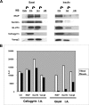
A, immunoadsorption of GLUT4-enriched sucrose gradient fractions show two distinction populations of GLUT4 vesicles. Separately pooled (as in Fig. 1) fractions for basal and insulin-treated rat adipocytes were precleared with anti-mouse IgG beads for 2 h, and the supernatants were immunoadsorbed with anti-cellugyrin (CG) beads for 2 h. Supernatants were collected and subjected to a subsequent GLUT4 (G4) immunoadsorption. All beads were washed three times with PBS, pH 7.4, and eluted with 100 μl of electrophoresis buffer. Equal proportions of IgG, cellugyrin, GLUT4, and supernatant were loaded on an SDS-polyacrylamide gel and analyzed by Western blot (WB) with proteins indicated as described under “Experimental Procedures.” B, quantification of Western blots reveals GLUT4 vesicles depleted of cellugyrin translocate upon insulin stimulation. Relative amounts of cellugyrin (CG), IRAP, GLUT4, and Vamp2 were determined by scanning and analysis with NIH image software, and the results are represented graphically. These results are representative of experiments done five times. A.U., arbitrary units; I.A., immunoadsorption.
FIGURE 3.
FIGURE 3.
Classes of proteins identified in precursor GSV and GSV compartments. Proteins found were categorized according to their presumed function as indicated in the pie chart (see supplemental Tables S1 and S2 for a complete list of all proteins identified). Details are given for presumptive cargo proteins in the table part of the figure as some of these are known GSV components (indicated by *). Relative protein abundance between the various immunoadsorption conditions were calculated by PAI as described (69). This index is based on the ratio of actual tryptic peptides identified by mass spectrometry divided by the theoretical tryptic peptides index within the mass range of 700–2600 Da, and the higher the number, the more abundant the protein. The table shows data from one of five similar experiments.
FIGURE 4.
FIGURE 4.
A, LM sucrose velocity gradient of LRP1 shows a similar sedimentation pattern to other GSV cargo. Sucrose sedimentation gradients were performed as described in Fig. 1 except for the Western blotting (W.B.) when a 4% SDS-PAGE was used to resolve the proteins prior to transfer. The chaperone protein BAP31 is shown as a loading control. B, insulin-stimulated translocation of LRP1 and GLUT4 to the PM. Fractionation of rat epididymal adipose tissue was performed as described under “Experimental Procedures.” PM (10 μg) was used for Western blotting of the indicated proteins, which were detected as in previous figures.
FIGURE 5.
FIGURE 5.
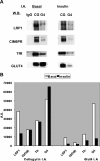
A, immunoadsorption of precursor GSVs and GSVs reveals CIM6PR, TfR, and LRP1 translocate upon insulin stimulation. Pooled sucrose gradient fractions were sequentially immunoadsorbed (I.A.) as described under “Experimental Procedures” and blotted for the proteins indicated as in previous figures. The results are representative of three independent experiments. B, Western blot (W.B.) quantitation of immunoadsorptions. The relative amounts of LRP1, IGFIIR, TfR, and GLUT4 (G4) were quantified by scanning and analysis with NIH image software and represented graphically. CG, cellugyrin; A.U., arbitrary units.
FIGURE 6.
FIGURE 6.
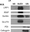
Reversible cross-linking of LM and immunoprecipitation (I.P.) by GLUT4 reveals a direct interaction between GSV proteins LRP1, IRAP, sortilin, and GLUT4. Primary rat adipocytes were fractionated as described under “Experimental Procedures.” A modified version (70) of cross-linking was performed. The isolated LM fraction (100 μg of protein) from rat epididymal fat was resuspended in 500 μl of PBS containing both protease and phosphatase inhibitors, and dithiobis(succinimidyl propionate) was added to a final concentration of 2 mm for 30 min at 20 °C. The samples were processed as described under “Experimental Procedures” and resolved by SDS-PAGE, transferred, and blotted for the indicated proteins. Detection was by ECL. PDI, protein-disulfide isomerase; W.B., Western blot; U.B., unbound.
FIGURE 7.
FIGURE 7.
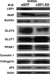
LRP1 depletion in 3T3-L1 adipocytes show decreased expression of IRAP, sortilin, and GLUT4. LRP1 and control eGFP stable knockdown 3T3-L1 fibroblasts were differentiated as described under “Experimental Procedures.” Cells were harvested, and whole cell extracts were prepared as described under “Experimental Procedures.” Protein (50 μg) was resolved by SDS-PAGE and Western blotted (W.B.) for the proteins shown. Detection was by enhanced chemiluminescence, and a representative blot is depicted. PPAR, peroxisome proliferator-activated receptor.
FIGURE 8.
FIGURE 8.
LRP1-depleted 3T3-L1 adipocytes show a decrease in insulin-stimulated 2-[3H]deoxyglucose uptake. Assays were performed in 6-well plates as described under “Experimental Procedures.” 2-[3H]Deoxyglucose (2-DG) counts were normalized to basal eGFP-transfected cells, and fold insulin-stimulation was assessed for each cell line. The results are presented as the mean ± S.D. for averaged duplicates from three separate experiments (# denotes p ≤ 0.05 and eGFP versus LRP1 shRNA).
FIGURE 9.
FIGURE 9.
Epididymal adipose tissues from LRP1 adipose-specific knock-out (KO) mice show decreased expression of GLUT4 and sortilin. Epididymal adipose tissues were isolated from 8-week-old male LRP1 lox/lox mice and an 8-week-old male aP2Cre+, LRP1 lox/lox mice. Cell lysates from the adipose tissues were prepared as described under “Experimental Procedures.” Proteins (25 μg) were resolved by SDS-PAGE, followed by Western blotting for the indicated proteins. The GLUT4 data were confirmed in a second animal pair (data not shown). WT, wild type; GAPDH, glyceraldehyde-3-phosphate dehydrogenase.
FIGURE 10.
FIGURE 10.
GST pulldowns containing the cytosolic domains of IRAP, sortilin, and LRP1 reveal its interactions with P115, and AS160. GST plasmids were constructed, induced, and purified as described under “Experimental Procedures.” Rat adipocytes were treated with 100 nm insulin or not and fractionated as described under “Experimental Procedures.” The cytosolic fraction (1 mg) from rat adipocytes stimulated (I) or not (B) with 100 nm insulin were precipitated with GST (C), GST-IRAP fusion protein (IRAP), GST-LRP1 fusion protein (LRP), and GST-sortilin fusion protein (sortilin) as described under “Experimental Procedures.” Pulldowns were subjected to SDS-PAGE, transferred onto a polyvinylidene difluoride membrane, and Western blotted (W.B.) for the indicated proteins.

References

    1. Huang S., Czech M. P. (2007) Cell Metab. 5, 237–252 - PubMed
    1. Zorzano A., Wilkinson W., Kotliar N., Thoidis G., Wadzinkski B. E., Ruoho A. E., Pilch P. F. (1989) J. Biol. Chem. 264, 12358–12363 - PubMed
    1. James D. E., Brown R., Navarro J., Pilch P. F. (1988) Nature 333, 183–185 - PubMed
    1. Cushman S. W., Wardzala L. J. (1980) J. Biol. Chem. 255, 4758–4762 - PubMed
    1. Holman G. D., Kozka I. J., Clark A. E., Flower C. J., Saltis J., Habberfield A. D., Simpson I. A., Cushman S. W. (1990) J. Biol. Chem. 265, 18172–18179 - PubMed

Publication types

MeSH terms