. 2010 Jan 1;285(1):392-400.
doi: 10.1074/jbc.M109.059055. Epub 2009 Oct 28.

Beta-catenin up-regulates Atoh1 expression in neural progenitor cells by interaction with an Atoh1 3' enhancer

Affiliations

Beta-catenin up-regulates Atoh1 expression in neural progenitor cells by interaction with an Atoh1 3' enhancer

Fuxin Shi et al. J Biol Chem. .

Abstract

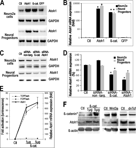
Atoh1, a basic helix-loop-helix transcription factor, plays a critical role in the differentiation of several epithelial and neural cell types. We found that beta-catenin, the key mediator of the canonical Wnt pathway, increased expression of Atoh1 in mouse neuroblastoma cells and neural progenitor cells, and baseline Atoh1 expression was decreased by siRNA directed at beta-catenin. The up-regulation of Atoh1 was caused by an interaction of beta-catenin with the Atoh1 enhancer that could be demonstrated by chromatin immunoprecipitation. We found that two putative Tcf-Lef sites in the 3' enhancer of the Atoh1 gene displayed an affinity for beta-catenin and were critical for the activation of Atoh1 transcription because mutation of either site decreased expression of a reporter gene downstream of the enhancer. Tcf-Lef co-activators were found in the complex that bound to these sites in the DNA together with beta-catenin. Inhibition of Notch signaling, which has previously been shown to induce bHLH transcription factor expression, increased beta-catenin expression in progenitor cells of the nervous system. Because this could be a mechanism for up-regulation of Atoh1 after inhibition of Notch, we tested whether siRNA to beta-catenin prevented the increase in Atoh1 and found that beta-catenin expression was required for increased expression of Atoh1 after Notch inhibition.


Figures

FIGURE 1.
FIGURE 1.
β-Catenin overexpression increased Atoh1 mRNA levels, and β-catenin silencing decreased Atoh1 mRNA levels. A, analysis of Atoh1 mRNA expression by RT-PCR showed that transfection of β-catenin (β-cat) into both Neuro2a cells and neural progenitors increased Atoh1 mRNA compared with untransfected cells (Ctl), whereas GFP transfection did not increase Atoh1 mRNA. Atoh1 transfection (Atoh1) was used as a positive control, and GAPDH was used as an internal control. B, increase in Atoh1 expression was quantified by real-time PCR in Neuro2a cells and neural progenitors from two independent experiments (each experiment in triplicate). The cells were transfected with either β-catenin (β-cat.) or Atoh1 (Atoh1) as positive controls or GFP (GFP) as a negative control. Atoh1 levels are expressed relative to untreated control cells (Ctl) and normalized to S18, a housekeeping gene. The increase in Atoh1 expression relative to the control was significant for both cell types transfected with β-catenin or Atoh1 (marked by asterisk). C, Atoh1 mRNA expression was analyzed by RT-PCR in Neuro2a cells and neural progenitors treated with siRNA. Atoh1 expression was decreased in the cells treated with β-catenin siRNA (siRNA-β-cat.) compared with non-targeting siRNA (siRNA-non-targ.) or no siRNA (Ctl). Cells treated with Atoh1 siRNA (siRNA-Atoh1) were used as a positive control. D, decrease in Atoh1 expression was quantified by real-time PCR from two independent experiments (each experiment in triplicate). The cells were transfected with β-catenin siRNA (siRNA-β-cat) or non-targeting siRNA (siRNA-non-targ). Atoh1 levels are expressed relative to untreated control cells (Ctl) and normalized to S18. Significant decreases in expression of Atoh1 are indicated by asterisks. E, activation of Tcf-Lef-mediated transcription was measured by TOPFlash luciferase reporter, and increased Atoh1 expression was quantified by real-time RT-PCR. Transfection of β-catenin (1 μg/ml or 5 μg/ml) increased TOPFlash activity (TOPFlash, dashed line) and Atoh1 mRNA expression in neural progenitor cells. FOPFlash (FOPFlash, dotted line), which contains mutant Tcf/Lef-binding sites remained unchanged in the cells transfected with β-catenin. F, nuclear fraction of unphosphorylated β-catenin and Atoh1 was examined by Western blotting in neural progenitors transfected with β-catenin, treated with Wnt3a, or transfected with dominant-negative Tcf4 (dnTcf). Overexpression of β-catenin increased the level of activated nuclear β-catenin (β-catenin*) and Atoh1. Wnt3a-conditioned medium compared with control conditioned medium (Ctl) also increased the level of active nuclear β-catenin and Atoh1. Conversely, overexpression of dominant-negative Tcf4 decreased the level of Atoh1.
FIGURE 2.
FIGURE 2.
Direct binding of β-catenin to the Atoh1 enhancer through Tcf-Lef. Binding of β-catenin to the Atoh1 enhancer was examined by ChIP. Chromatin was cross-linked in Neuro2a cells followed by precipitation with nonimmune IgG, β-catenin antibody, or Tcf-Lef antibody. The sheared Atoh1 enhancer fragments were amplified from the precipitated chromatin. The primers for DNA at the two ends of the Atoh1 enhancer, including the predicted Tcf-Lef binding sites (148–434 and 928–1123) and adjacent fragments, were found to amplify DNA from the immunoprecipitate. In a control, chromatin precipitated with nonimmune IgG (serum) did not contain DNA that could be amplified with these primers. Input refers to DNA without antibody precipitation. The extent of amplification of the fragments varied as a function of the extent of shearing of the chromatin.
FIGURE 3.
FIGURE 3.
β-Catenin interacts with the Atoh1 3′ enhancer in a complex with Tcf-Lef. A, nuclear lysate from Neuro2a cells was incubated with biotin-labeled DNA probes corresponding to enhancer sequence 297–326 (probe 309) and 956–985 (probe 966), which contained two predicted β-catenin/Tcf-Lef binding sequences, and Atoh1 enhancer-binding proteins were collected with magnetic beads coupled to streptavidin. Proteins were analyzed by Western blotting using antibodies specific for β-catenin and Tcf-Lef. β-Catenin (β-catenin) was found in the bead-eluted proteins (probe 309 and probe 966), indicating an interaction with the probes; binding was inhibited by competition with excess unlabeled probes (comp 309 and comp 966). Binding was inhibited when predicted β-catenin/Tcf-Lef binding sequences were mutated in the probes (mutant 309 and mutant 966). Tcf-Lef was also found in this fraction (Tcf/Lef) indicating formation of a complex involving Tcf-Lef factors and β-catenin. B, expression of Atoh1 in untransfected Neuro2a cells and neural progenitors (Ctl) was increased in the same cells transfected with 1 μg/ml β-catenin (β-cat). The level of Atoh1 was decreased in a dose-dependent manner when cells were co-transfected with dominant-negative Tcf4 (β-cat + dn Tcf). The Atoh1 levels were analyzed by real-time PCR in two independent experiments (each experiment in triplicate). The asterisks mark significant differences in Atoh1 expression compared with control.
FIGURE 4.
FIGURE 4.
β-Catenin binding to the Atoh1 enhancer at a Tcf-Lef binding site accounts for the functional effect of β-catenin on Atoh1 expression. Atoh1 enhancer was used to drive luciferase expression in the pGL-3 reporter vector (Atoh1-Luc). The β-catenin binding sites on the Atoh1 enhancer were mutated individually (mutant 309; mutant 966) or both binding sites were mutated (2X mutant). The reporter construct or mutant reporters were co-transfected into Neuro2a cells with overexpression of β-catenin. Overexpression of β-catenin (black bars) activated reporter expression up to 4-fold (Atoh1-Luc), whereas β-catenin overexpression had no effect on expression of the pGL-3 construct in the absence of the Atoh1 enhancer (Luc, gray bars). Mutant 309 (Atoh1-Luc, mutant 309) decreased β-catenin-mediated activation of the enhancer 53% (from 3.9–1.84-fold), whereas mutant 966 (Atoh1-Luc, mutant 966) decreased activation of the enhancer 26% (from 3.9–2.91-fold). Each treatment was in triplicate, and the values shown are from two independent experiments with significant changes indicated by an asterisk (compared with the unmutated Atoh1 reporter). The double mutation (Atoh1, 2X mutant) completely abolished activation of the enhancer. Neither single nor double mutation in the absence of β-catenin had an effect on reporter expression (gray bars).
FIGURE 5.
FIGURE 5.
Increased expression of Atoh1 after Notch inhibition is partly caused by β-catenin expression. A, to observe the effect of Notch inhibition we assessed expression of β-catenin and Atoh1 in bone marrow-derived MSCs by Western blot. The expression of β-catenin was increased in MSCs treated with a γ-secretase inhibitor, DAPT, at 10 μm and 50 μm. Atoh1 expression was also increased (Atoh1). Treatment of the cells with GSK3β inhibitor (GSKi) also increased the expression of β-catenin and Atoh1. B, effect of two siRNAs to β-catenin (siRNA-β-cateninA; siRNA-β-cateninB) was evaluated by quantitative RT-PCR. Treatment with the siRNAs decreased the expression of β-catenin in a dose-dependent manner; non-targeting siRNA (siRNA-non-targeting) had no significant effect. C, to determine whether increased expression of Atoh1 seen after Notch inhibition was related to β-catenin expression the levels of β-catenin and Atoh1 were examined by Western blot. The elevated β-catenin after DAPT treatment (β-catenin) was decreased by siRNA to β-catenin. The increased expression of Atoh1 with DAPT could also be decreased by blocking β-catenin expression with the siRNA but not with non-targeting siRNA. D, we tested the effect of disrupting the Notch pathway using Pofut1−/− cells that have a mutation that prevents Notch signaling. Atoh1 expression was higher in these cells and the γ-secretase inhibitor could not further increase Atoh1 expression as determined by quantitative RT-PCR (pofut-), compared with wild-type cells (Rosa 26 and pofut+). The deceased level of β-catenin using siRNA (siRNA-β-catenin) resulted in a decrease in expression of Atoh1 (pofut-), confirming that β-catenin signaling under conditions of decreased Notch signaling was partly responsible for the increased level of Atoh1. The decrease in Atoh1 expression in cells treated with β-catenin siRNA was significant compared with the control and DAPT treatment. E, inhibition of Notch activity with DAPT (DAPT) increased activated β-catenin (β-catenin*) and Atoh1 expression. Disruption of β-catenin-mediated transcription by overexpression of dominant-negative Tcf4 (dn Tcf) reversed the increase of Atoh1 expression in cells treated with the Notch inhibitor. Overexpression of Notch intracellular domain (Notch) decreased activated β-catenin and Atoh1 expression. Activation of β-catenin-mediated transcription by Wnt3a rescued the decrease of Atoh1 expression in cells with elevation of Notch activity. F, inhibition of Notch with DAPT decreased phosphorylated GSK3β (GSK3β*Y216), increased activated β-catenin (β-catenin*), and increased Atoh1 expression. Conversely, activation of Notch intracellular domain increased phosphorylated GSK3β, decreased activated β-catenin, and reduced Atoh1 expression. The total GSK3β remained unchanged.
FIGURE 6.
FIGURE 6.
Schematic diagram illustrates regulation of Atoh1 by β-catenin. The positive sign and arrow represent the increased transcription of Atoh1 upon binding of β-catenin-Tcf/Lef to the Atoh1 3′-enhancer. The resulting Atoh1 acts to up-regulate its own expression by binding to the same enhancer. Stimulation by β-catenin accounts for the up-regulation of Atoh1 after Notch inhibition. Atoh1 levels are determined by negative regulation by transcription factors such as Hes1 and 5 when Notch signaling is active.

References

    1. Bermingham N. A., Hassan B. A., Price S. D., Vollrath M. A., Ben-Arie N., Eatock R. A., Bellen H. J., Lysakowski A., Zoghbi H. Y. (1999) Science 284, 1837–1841 - PubMed
    1. Yang Q., Bermingham N. A., Finegold M. J., Zoghbi H. Y. (2001) Science 294, 2155–2158 - PubMed
    1. Bertrand N., Castro D. S., Guillemot F. (2002) Nat. Rev. Neurosci. 3, 517–530 - PubMed
    1. Hirata H., Tomita K., Bessho Y., Kageyama R. (2001) EMBO J. 20, 4454–4466 - PMC - PubMed
    1. Ross S. E., Greenberg M. E., Stiles C. D. (2003) Neuron 39, 13–25 - PubMed

Publication types

MeSH terms