

Progressive chondrocyte death after impact injury indicates a need for chondroprotective therapy
- PMID: 19864505
- PMCID: PMC3425608
- DOI: 10.1177/0363546509348840
Progressive chondrocyte death after impact injury indicates a need for chondroprotective therapy
Abstract
Background: Impact injury to articular cartilage can lead to posttraumatic osteoarthritis.
Hypotheses: This study tests the hypotheses that (1) chondrocyte injury occurs after impact at energies insufficient to fracture the cartilage surface, and that (2) cartilage injury patterns vary with impact energy, time after injury, and cartilage thickness.
Study design: Controlled laboratory study.
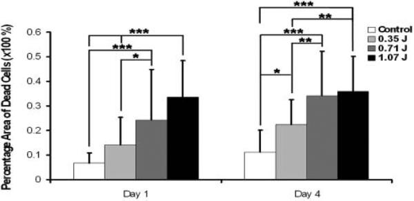
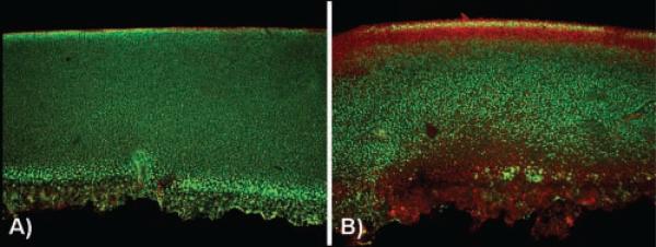
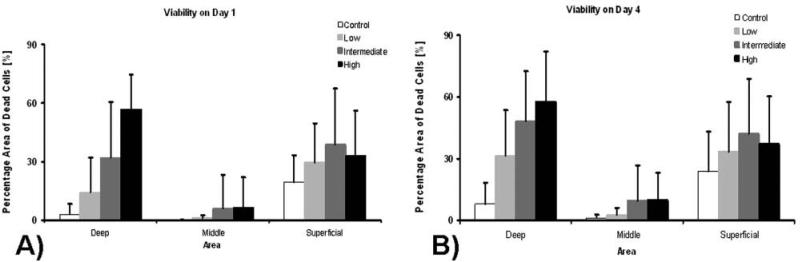
Methods: Fresh bovine osteochondral cores were randomly divided into 5 groups: (1) control, (2) 0.35 J, (3) 0.71 J, (4) 1.07 J, and (5) 1.43 J impact energies. Cores were subjected to computer-controlled impact loading and full-thickness sections were then prepared and incubated in Dulbecco's Modified Eagle's Medium/F12 at 37 degrees C. Adjacent sections were harvested 1 and 4 days after impact for viability staining and fluorescent imaging. The area of dead and living chondrocytes was quantified using custom image analysis software and reported as a percentage of total cartilage area.
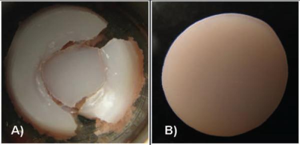
Results: The highest impact energy fractured the cartilage in all cores (1.43 J, n = 17). Seventy-three percent and 64% of the osteochondral cores remained intact after lower energy impacts of 0.71 J and 1.07 J, respectively. At lower energy levels, fractured cores were thinner (P <.01) than those remaining intact. In cores remaining intact after impact injury, chondrocyte death increased with increasing impact energy (P <.05) and with greater time after impact (P <.05). A progressive increase in dead cells near the bone/cartilage interface and at the articular surface was observed.
Conclusion: These data showing progressive chondrocyte death after impact injury at energies insufficient to fracture the cartilage surface demonstrate a potential need for early chondroprotective therapy.
Clinical relevance: These data show that efforts to reduce chondrocyte morbidity after joint injury may be a useful strategy to delay or prevent the onset of posttraumatic osteoarthritis.
Figures
Comment in
-
Ben would approve.Am J Sports Med. 2010 Aug;38(8):NP2-3. doi: 10.1177/0363546510371673. Am J Sports Med. 2010. PMID: 20675647 No abstract available.
References
-
- Baars DC, Rundell SA, Haut RC. Treatment with the non-ionic surfactant poloxamer P188 reduces DNA fragmentation in cells from bovine chondral explants exposed to injurious unconfined compression. Biomech Model Mechanobiol. 2006;5(2-3):133–139. - PubMed
-
- Borrelli J, Jr, Ricci WM. Acute effects of cartilage impact. Clin Orthop Relat Res. 2004;423:33–39. - PubMed
-
- Borrelli J, Jr, Silva MJ, Zaegel MA, Franz C, Sandell LJ. Single high-energy impact load causes posttraumatic OA in young rabbits via a decrease in cellular metabolism. J Orthop Res. 2009;27(3):347–352. - PubMed
-
- Borrelli J, Jr, Tinsley K, Ricci WM, Burns M, Karl IE, Hotchkiss R. Induction of chondrocyte apoptosis following impact load. J Orthop Trauma. 2003;17(9):635–641. - PubMed
-
- Brown TD, Johnston RC, Saltzman CL, Marsh JL, Buckwalter JA. Posttraumatic osteoarthritis: a first estimate of incidence, prevalence, and burden of disease. J Orthop Trauma. 2006;20(10):739–744. - PubMed
Publication types
MeSH terms
Grants and funding
LinkOut - more resources
Full Text Sources
Other Literature Sources
Medical
Miscellaneous
