. 2009 Oct 28;29(43):13435-44.
doi: 10.1523/JNEUROSCI.3257-09.2009.

Identification of two distinct macrophage subsets with divergent effects causing either neurotoxicity or regeneration in the injured mouse spinal cord

Affiliations

Identification of two distinct macrophage subsets with divergent effects causing either neurotoxicity or regeneration in the injured mouse spinal cord

Kristina A Kigerl et al. J Neurosci. .

Abstract

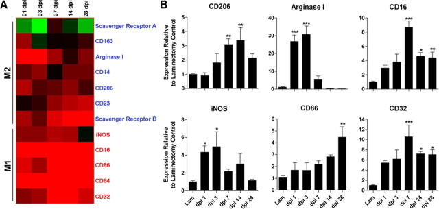
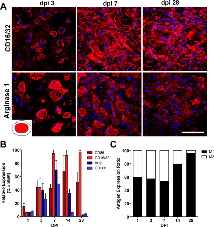
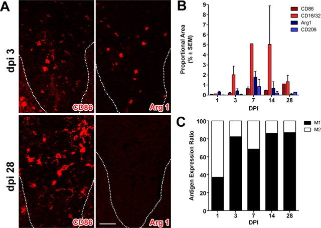
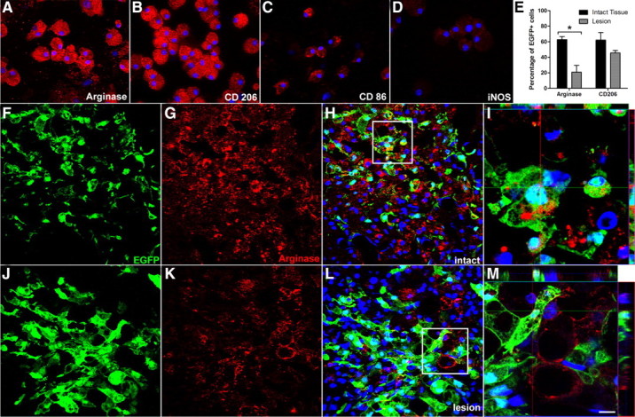
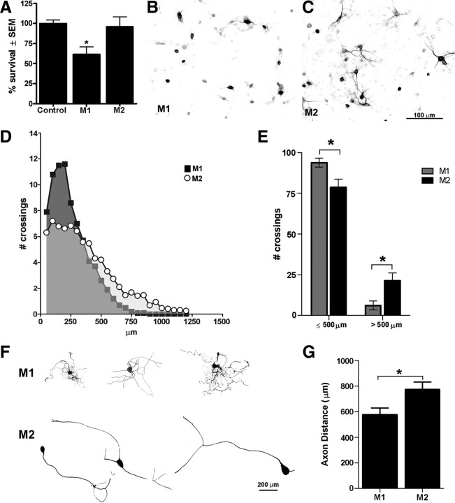
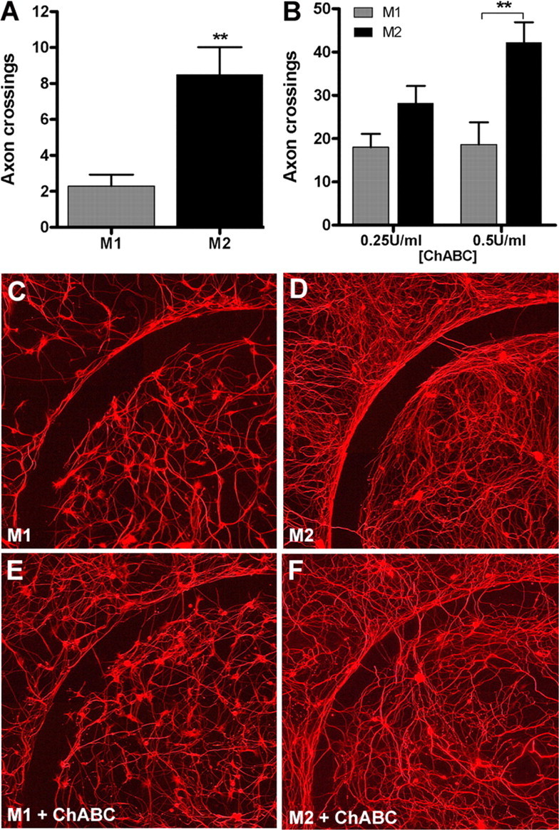
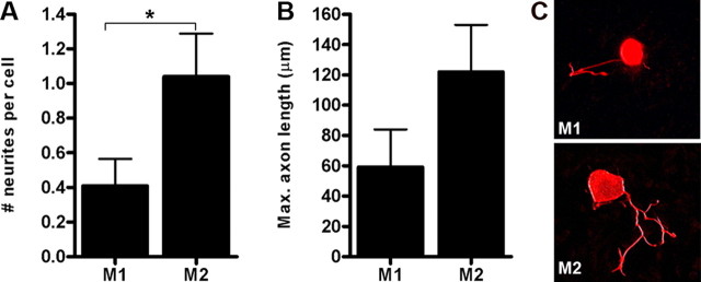
Macrophages dominate sites of CNS injury in which they promote both injury and repair. These divergent effects may be caused by distinct macrophage subsets, i.e., "classically activated" proinflammatory (M1) or "alternatively activated" anti-inflammatory (M2) cells. Here, we show that an M1 macrophage response is rapidly induced and then maintained at sites of traumatic spinal cord injury and that this response overwhelms a comparatively smaller and transient M2 macrophage response. The high M1/M2 macrophage ratio has significant implications for CNS repair. Indeed, we present novel data showing that only M1 macrophages are neurotoxic and M2 macrophages promote a regenerative growth response in adult sensory axons, even in the context of inhibitory substrates that dominate sites of CNS injury (e.g., proteoglycans and myelin). Together, these data suggest that polarizing the differentiation of resident microglia and infiltrating blood monocytes toward an M2 or "alternatively" activated macrophage phenotype could promote CNS repair while limiting secondary inflammatory-mediated injury.


Figures

Figure 1.
Figure 1.
Spinal cord injury induces changes in the expression of genes associated with M1 and M2 macrophages. A, cDNA microarray “heat map” showing clustering of M1 and M2 genes as a function of time after injury. All genes shown are upregulated or downregulated more than twofold compared with uninjured controls. Black indicates no change in gene expression, whereas green or red indicate a decrease or increase in expression, respectively, relative to uninjured control samples. Note transient induction of M2 genes in contrast with early and sustained induction of M1 genes. B, Quantitative real-time PCR confirms select M1 and M2 gene expression changes noted via microarray (n = 4 per time for microarray; n = 3–5 per time for PCR). (ANOVA, p < 0.01 for all genes; *p < 0.05, **p < 0.01, ***p < 0.001 vs laminectomy control.).
Figure 2.
Figure 2.
Macrophages with an M1 phenotype dominate sites of spinal cord injury. A, CD16/32+ M1 macrophages and arginase 1+ M2 macrophages coexist at the lesion epicenter during the first week after injury; however, only M1 macrophages persist until 28 dpi. B, Quantitation of macrophages expressing M1 and M2 phenotypic markers as a function of time after SCI. C, When expressed as a ratio of M1/M2 cells, there is an obvious shift toward an M1 macrophage phenotype after the first week after injury. [M1 and M2 markers; red, AF546 and nuclear stain with 4′,6′-diamidino-2-phenylindole (DAPI); blue]. Scale bar, 20 μm.
Figure 3.
Figure 3.
M1 macrophages dominate zones of Wallerian degeneration after SCI. A, Similar to the macrophage response that evolves at the lesion epicenter (see Fig. 2), M1 (CD86+) and M2 (Arg 1) macrophages coexist within the dorsal funiculus at 3 dpi, but only M1 macrophages persist until 28 dpi (dotted line indicates the border of the gray matter and dorsal funiculus). B, Quantitation of macrophages expressing M1 and M2 markers in the dorsal funiculus at different times after SCI. C, When expressed as a ratio of M1/M2 cells, there is an obvious shift toward an M1 macrophage by 3 dpi. (M1 and M2 markers; red, AF546 and nuclear stain with DAPI; blue). Scale bar, 20 μm.
Figure 4.
Figure 4.
The M2 macrophage phenotype is downregulated in the injured spinal cord. The phenotype of EGFP+ M2 macrophages was confirmed ex vivo (A–D), and then cells were microinjected into areas of intact (F–I) or injured (7 dpi; J–M) spinal cord. The percentage of EGFP+ cells expressing arginase 1 (or CD206) (supplemental Fig. 4, available at www.jneurosci.org as supplemental material) was reduced when injected into injured spinal cord (n = 3 per group; *p < 0.05 vs intact for arginase 1) (E). Confocal images of EGFP+ cells stained with antibodies labeling the M2 marker arginase 1 (red) reveal SCI-dependent reduction in the M2 phenotype (compare G–I with K–M). I, M, High-powered images of boxed areas in H and L, respectively. Notice loss of arginase 1 labeling inside EGFP+ macrophages in injured spinal cord. Instead, a small population of EGFP-negative parenchymal cells express low levels of arginase 1. Scale bar: F–H, J–L, 20 μm; I, 7.5 μm; M, 5.5 μm.
Figure 5.
Figure 5.
M1 and M2 macrophages exert disparate effects on neuron survival and axon growth. M1 macrophages are neurotoxic in vitro (A–C). Regardless of whether cortical neuron survival (7 d in vitro) was assessed in a media transfer assay (A) or in response to M1 or M2 macrophages in a trans-well system (B, C), M1 macrophages were neurotoxic. Cortical neuron survival was quantified using a MAP2 ELISA (A; *p < 0.05 vs control and M2 MCM). M2 macrophages had no adverse effect on neurons (A, C). MCM from M1 and M2 cells also trigger distinct neurite outgrowth patterns in DRG neurons (D–G). Sholl analysis was used to measure neurite outgrowth length and complexity (D). M1 MCM stimulated a short arborizing growth pattern with axons usually terminating within 500 μm of the cell soma (D–G). Conversely, M2 MCM induced long unipolar or bipolar axonal extensions with less branching evident near the soma and overall length often exceeding 1 mm (D–G) (*p < 0.05 for E, G; data are representative of 2 independent experiments; n = 15–20 DRGs analyzed).
Figure 6.
Figure 6.
M2 macrophages promote axon growth across an inhibitory CSPG gradient. Adult DRG neurons were treated with M1 or M2 MCM for 5 d in vitro, and then axons crossing over an aggrecan barrier were quantified. M2 MCM consistently enhanced DRG axon crossings compared with M1 MCM (p < 0.01; A, C, D). M2 MCM-mediated axon growth was augmented in the presence of increasing concentrations of chABC (**p < 0.01 at 0.5 U/ml chABC vs M1 MCM; B, E, F). Data are representative of three independent experiments; n = 12 spots per condition.
Figure 7.
Figure 7.
M2 macrophages enhance axon growth on an inhibitory myelin (MAG) substrate. DRG neurons plated on a monolayer of CHO cells engineered to express MAG (Domeniconi et al., 2002) were treated with M1 or M2 MCM for 48 h, and then neurite outgrowth was assessed. M2 MCM increased the number (A) and length (B) of neurites extending from DRG neurons (A; *p < 0.05). Representative β-tubulin III+ DRG neurons are provided showing the median number and length of neurites elicited by M1 or M2 MCM (C). These data represent mean of n = 20–25 neurons analyzed from an individual experiment.

References

    1. Auffray C, Fogg D, Garfa M, Elain G, Join-Lambert O, Kayal S, Sarnacki S, Cumano A, Lauvau G, Geissmann F. Monitoring of blood vessels and tissues by a population of monocytes with patrolling behavior. Science. 2007;317:666–670. - PubMed
    1. Barrette B, Hébert MA, Filali M, Lafortune K, Vallières N, Gowing G, Julien JP, Lacroix S. Requirement of myeloid cells for axon regeneration. J Neurosci. 2008;28:9363–9376. - PMC - PubMed
    1. Bethea JR, Nagashima H, Acosta MC, Briceno C, Gomez F, Marcillo AE, Loor K, Green J, Dietrich WD. Systemically administered interleukin-10 reduces tumor necrosis factor-alpha production and significantly improves functional recovery following traumatic spinal cord injury in rats. J Neurotrauma. 1999;16:851–863. - PubMed
    1. Blight AR. Effects of silica on the outcome from experimental spinal cord injury: implication of macrophages in secondary tissue damage. Neuroscience. 1994;60:263–273. - PubMed
    1. Block ML, Zecca L, Hong JS. Microglia-mediated neurotoxicity: uncovering the molecular mechanisms. Nat Rev Neurosci. 2007;8:57–69. - PubMed

Publication types

MeSH terms

Substances