. 2009 Oct 28;29(43):13720-9.
doi: 10.1523/JNEUROSCI.3018-09.2009.

Control of cortical axon elongation by a GABA-driven Ca2+/calmodulin-dependent protein kinase cascade

Affiliations

Control of cortical axon elongation by a GABA-driven Ca2+/calmodulin-dependent protein kinase cascade

Natsumi Ageta-Ishihara et al. J Neurosci. .

Abstract

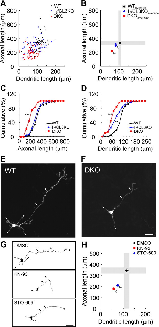
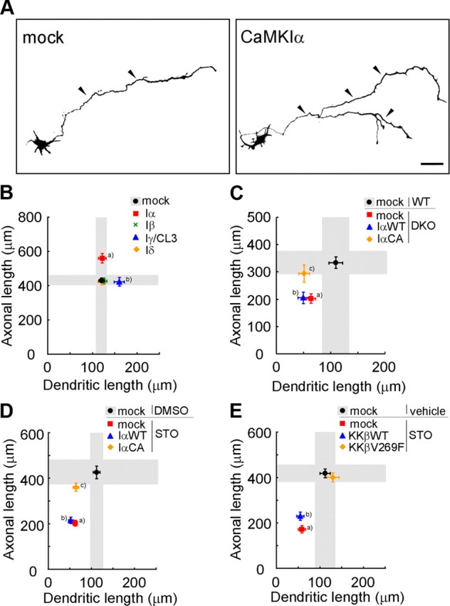
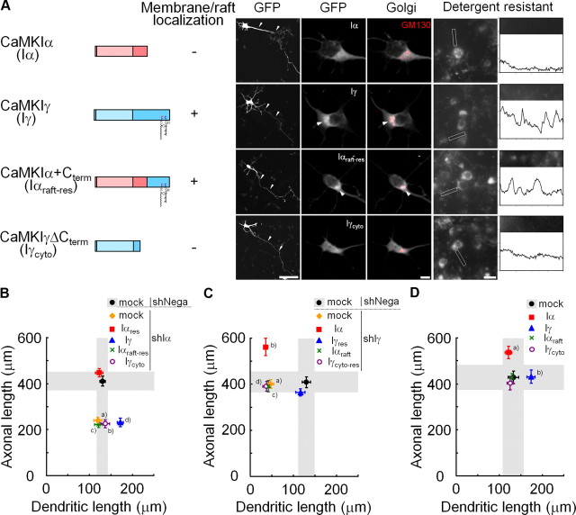
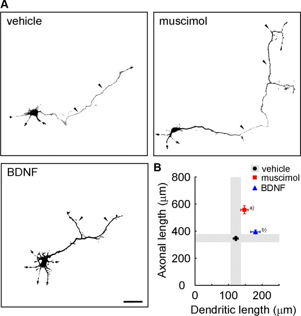
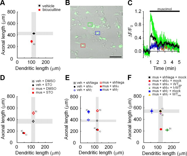
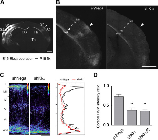
Ca(2+) signaling plays important roles during both axonal and dendritic growth. Yet whether and how Ca(2+) rises may trigger and contribute to the development of long-range cortical connections remains mostly unknown. Here, we demonstrate that two separate limbs of the Ca(2+)/calmodulin-dependent protein kinase kinase (CaMKK)-CaMKI cascades, CaMKK-CaMKIalpha and CaMKK-CaMKIgamma, critically coordinate axonal and dendritic morphogenesis of cortical neurons, respectively. The axon-specific morphological phenotype required a diffuse cytoplasmic localization and a strikingly alpha-isoform-specific kinase activity of CaMKI. Unexpectedly, treatment with muscimol, a GABA(A) receptor agonist, selectively stimulated elongation of axons but not of dendrites, and the CaMKK-CaMKIalpha cascade critically mediated this axonogenic effect. Consistent with these findings, during early brain development, in vivo knockdown of CaMKIalpha significantly impaired the terminal axonal extension and thereby perturbed the refinement of the interhemispheric callosal projections into the contralateral cortices. Our findings thus indicate a novel role for the GABA-driven CaMKK-CaMKIalpha cascade as a mechanism critical for accurate cortical axon pathfinding, an essential process that may contribute to fine-tuning the formation of interhemispheric connectivity during the perinatal development of the CNS.


Figures

Figure 1.
Figure 1.
CaMKK-dependent CaMK cascades control cortical axonal and dendritic growth. A, B, A scattered plot (orthogonal plot) of data points (A) and averages (B) for both axonal and dendritic lengths obtained of individual neurons. Black circles, Wild type (WT). Blue triangles, Iγ/CL3 knock-out (Iγ/CL3 KO). Red squares, CaMKKα/β-double knock-out (DKO). Number of neurons: WT, n = 52; Iγ/CL3 KO, n = 44; DKO, n = 52. aDendrite, p < 0.01; bAxon, p < 0.001; dendrite, p < 0.001 (one-way ANOVA with Tukey's test comparison with WT). C, D, Cumulative probability analysis for total axonal length (C) and total dendritic length (D) in neurons from WT, DKO, and Iγ/CL3 KO mice. Number of neurons: WT, n = 52; DKO, n = 52; Iγ/CL3 KO, n = 44. **p < 0.01; ***p < 0.001, Kolmogorov–Smirnov test comparison with WT. E, F, Cortical neurons (2 d in vitro) from CaMKKα/β-DKO mice (F) showed impaired growth of axons (arrowheads) and dendrites (arrows) compared with neurons from WT mice (E). Scale bar, 25 μm. G, Treatment with KN-93, a general CaMK inhibitor, and STO-609, a blocker of CaMKKα/β, the upstream kinases of all CaMKI/IV, from 6 to 48 h after plating impaired both axonal (arrowheads) and dendritic (arrows) growth. Scale bar, 50 μm. H, An orthogonal plot shows a quantitative analysis of axonal and dendritic morphometric parameters from each neuron. Number of neurons: DMSO, n = 48; KN-93, n = 43; STO-609, n = 48. a,bAxon, p < 0.001; dendrite, p < 0.001 (one-way ANOVA with Tukey's test comparison with DMSO).
Figure 2.
Figure 2.
Knockdown of CaMKIα specifically impairs axonal but not dendritic growth. A, Efficient downregulation of exogenous GFP-CaMKIα was achieved by a CaMKIα-targeted shRNA vector (shCaMKIα), but not by a control vector (shNega), in rat cortical neurons. The mRFP1 expression, which was driven by a dual promoter in a pSUPER+mRFP1 vector, remained unchanged. Scale bar, 50 μm. B, Knockdown of endogenous CaMKIα was evaluated by Western blot analysis using an anti-CaMKIα antibody. Rat cortical neurons were transfected with pSUPER-shNega or pSUPER-shCaMKIα by electroporation, and the cells were lysed at 2 DIV. shCaMKIα suppressed endogenous CaMKIα, whereas the control mRFP1 expression level remained unchanged. C, D, shCaMKIα-expressing rat cortical neurons (shCaMKIα) (D) showed impaired axonal growth (arrowheads), whereas the dendritic morphology was spared (arrows) compared with neurons from shNega-expressing rat cortical neurons (shNega) (C). Scale bar, 25 μm. E, An orthogonal plot of averaged data; n = 15 for all groups. aAxon, p < 0.001. bDendrite, p < 0.001 (one-way ANOVA with Tukey's test comparison with shNega). Scale bar, 25 μm. F, Introduction of shCaMKIα-resistant wild-type GFP-CaMKIα (WTres) successfully rescued the axonal defect elicited by shCaMKIα. In contrast, an shCaMKIα-resistant kinase-inactive GFP-CaMKIα (a K49Ares point mutant) was unable to rescue the shCaMKIα phenotype. n = 15 for all groups. a,bAxon, p < 0.001 (one-way ANOVA with Tukey's test comparison with shNega + mock).
Figure 3.
Figure 3.
A specific role for a CaMKK–CaMKIα cascade in promoting axonal growth in cortical neurons. A, Representative images of rat cortical neurons transfected with GFP-CaMKIα. Scale bar, 50 μm. B, Overexpression of CaMKIα and CaMKIγ facilitated axonal and dendritic growth, respectively. n = 15 for all groups. aAxon, p < 0.001; bdendrite, p < 0.05 (one-way ANOVA with Tukey's test comparison with mock). C, The axonal growth defect in DKO mice was selectively rescued by coexpression of a constitutively active CaMKIα (CaMKIαCA), but not by a wild-type CaMKIα (CaMKIαWT); the dendritic growth defect was left unaltered; n = 15 for all groups. aAxon, p < 0.01; dendrite, p < 0.05; baxon, p < 0.01; dendrite, p < 0.001; cdendrite, p < 0.01 (one-way ANOVA with Tukey's test comparison with WT plus mock). D, Only the axonal growth defects caused by STO-609 treatment were rescued by expression of a constitutively active CaMKIα (CaMKIαCA), but not of a wild-type CaMKIα (CaMKIαWT). Dendritic growth defects remained unchanged; n = 15 for all groups. a,bAxon, p < 0.001; dendrite, p < 0.001; cDendrite, p < 0.001 (one-way ANOVA with Tukey's test comparison with DMSO plus mock). E, Both axonal and dendritic growth defects caused by STO-609 treatment were rescued by introducing an STO-609-resistant CaMKKβ mutant (V269F), but not a CaMKKβ-WT. n = 15 for all groups. a,bAxon, p < 0.001; dendrite, p < 0.01 (one-way ANOVA with Tukey's test comparison with vehicle plus mock).
Figure 4.
Figure 4.
Functional segregation of CaMKK–CaMKIα and CaMKK–CaMKIγ cascades. A, The domain structures and subcellular localizations of CaMKIα (Iα), CaMKIγ/CL3 (Iγ), and their chimeras. GFP-CaMKIα (Iα), CaMKIγ/CL3 (Iγ), and their chimeras distribution detected by anti-GFP immunostaining showed colocalization with a Golgi marker, GM130. GFP-Iγ and Iαraft-res signals were also enriched within Golgi (arrowheads). Single representative confocal sections are shown for Golgi localization. GFP-Iγ and -Iαraft-res fluorescence was retained after detergent treatment in a punctate manner in 2 DIV cortical neurons along the dendrites, demonstrating a sizable portion of detergent-resistant GFP-Iγ and Iαraft-res in the dendritic rafts. Line scans of pixel fluorescence, performed within a chosen field of a 15 μm dendritic segment. Scale bars: right, 50 μm; middle, 5 μm; left, 100 μm. B, Neither Iγ, Iαraft-res, nor Iγcyto were able to rescue the axonal phenotype caused by knockdown of CaMKIα; n = 15 for all groups. a,b,cAxon, p < 0.001; daxon, p < 0.001, dendrite, p < 0.01 (one-way ANOVA with Tukey's test comparison with shNega plus mock). C, Neither Iα, Iαraft, nor Iγcyto-res were able to rescue the dendritic phenotype caused by knockdown of CaMKIγ/CL3; n = 15 for all groups. a,c,dDendrite, p < 0.001; baxon, p < 0.001, dendrite, p < 0.001 (one-way ANOVA with Tukey's test comparison with shNega plus mock). D, Overexpression of CaMKIα, specifically increased axon length in cortical neurons; n = 15 for all groups; n = 15 for all groups. aAxon, p < 0.05; bdendrite, p < 0.001 (one-way ANOVA with Tukey's test comparison with mock).
Figure 5.
Figure 5.
Muscimol, a GABAA receptor agonist, specifically stimulates elongation of axons in cultured cortical neurons. A, B, Representative images (A) and ensemble data (B) of immature cortical neurons treated with either muscimol (a GABAA receptor agonist) or BDNF. Muscimol significantly promoted axonal growth (arrowheads). In contrast, BDNF had no effect on axons but mainly affected dendrites. Scale bar, 50 μm. n = 15 for all groups. aAxon, p < 0.001; bdendrite, p < 0.01 (one-way ANOVA with Tukey's test comparison with vehicle).
Figure 6.
Figure 6.
Activation of GABAA receptors promotes axonal growth via the CaMKK–CaMKIα pathway in immature cortical neurons. A, Bicuculline, a GABAA receptor antagonist, blocked axonal growth; n = 15 for all groups. aAxon, p < 0.001 (Student's t test comparison with vehicle). B, Embryonic cortical neurons (1 DIV) were loaded with a calcium indicator, Fluo-4 AM, and calcium responses were measured by time-lapse imaging. A green fluorescence image was overlaid on a differential interference contrast image. The colored boxes indicate the location of cells shown in C. Scale bar, 50 μm. C, Representative calcium responses in individual cells after muscimol administration. Three different types of calcium responses were revealed (green, blue, and red). An averaged response from 10 cells in a microscopic field is revealed in black. D, Both basal and muscimol-stimulated axonal growths were suppressed with STO-609, a specific blocker of CaMKKα/β; n = 15 for all groups. Axon: two-way ANOVA, muscimol effect, F(1,56) = 14.38, p = 0.0004; drug effect, F(1,56) = 225.63, p < 0.0001; muscimol × drug, F(1,56) = 15.79, p = 0.0002. Dendrite: two-way ANOVA, muscimol effect, F(1,56) = 0.39, p = 0.5336; drug effect, F(1,56) = 105.76, p < 0.0001; muscimol × drug, F(1,56) = 0.13, p = 0.7199. a,bAxon, p < 0.001 (comparison with vehicle plus DMSO); caxon, p < 0.001 (comparison with muscimol plus DMSO); n.s. (comparison with vehicle plus STO). E, CaMKIα knockdown quantitatively inhibited axonal growth induced by muscimol treatment, to an extent similar to that obtained with STO-609; n = 15 for all groups. Axon: two-way ANOVA, muscimol effect, F(1,84) = 66.44, p < 0.0001; RNAi effect, F(2,84) = 168.04, p < 0.0001; muscimol × RNAi, F(2,84) = 19.96, p < 0.0001. Dendrite: two-way ANOVA, muscimol effect, F(1,84) = 0.23, p = 0.6305; RNAi effect, F(2.84) = 61.58, p < 0.0001; muscimol × RNAi, F(2.84) = 0.01, p = 0.9888. a,bAxon, p < 0.001 (comparison with vehicle plus shNega); caxon, p < 0.001 (comparison with muscimol plus shNega); n.s. (comparison with vehicle plus shIα); daxon, p < 0.001 (comparison with vehicle plus shIγ); n.s. (comparison with muscimol plus shNega). F, Introduction of shCaMKIα-resistant wild-type GFP-CaMKIα (WTres) specifically rescued the suppression of muscimol-induced axonal growth triggered by knockdown of CaMKIα; n = 15 for all groups. aAxon, p < 0.001; dendrite, n.s.; baxon, n.s.; dendrite, p < 0.001 (one-way ANOVA with Tukey's test comparison with muscimol plus shIα plus mock); caxon, n.s.; dendrite, p < 0.001 (t test comparison with muscimol plus shIγ plus mock).
Figure 7.
Figure 7.
Knockdown of CaMKIα impairs terminal extension of callosal axons in vivo. A, A control coronal section was obtained near the posterior end of the corpus callosum, from a P16 pup electroporated in utero with pSUPER-shNega and pCAG-EGFP on E15.5. The somatodendritic regions of layer II/III neurons were strongly labeled (asterisks) in the somatosensory cortex, from which callosal axons projected toward the contralateral cortical areas at the S1/S2 border region (arrowhead). Scale bar, 1 mm. CC, Corpus callosum; Hi, hippocampus; Th, thalamus. B, Terminal extension of callosal axons into the contralateral cortical layers was severely disrupted in CaMKIα-knockdown neurons (shKIα), although axons were able to reach the white matter (WM) beneath S1/S2 area (arrowhead). Scale bar, 1 mm. C, Axonal extension and terminal branch arborization were strongly impaired in layers II/III in CaMKIα-knockdown neurons, as illustrated by the magnified images of GFP marker showing the total axonal volumes present in the cortical layers (in pseudocolor), or by a one-dimensional fluorescence intensity profile analysis. Scale bar, 200 μm. D, Quantification of the cortical wiring defect caused by an aberrant terminal axon extension in the cortex. Two independent RNAi constructs (shKIα and shKIα #2) gave similar results. **p < 0.01 (one-way ANOVA with Tukey's test comparison with shNega).

Comment in

References

    1. Behar TN, Li YX, Tran HT, Ma W, Dunlap V, Scott C, Barker JL. GABA stimulates chemotaxis and chemokinesis of embryonic cortical neurons via calcium-dependent mechanisms. J Neurosci. 1996;16:1808–1818. - PMC - PubMed
    1. Behar TN, Schaffner AE, Scott CA, O'Connell C, Barker JL. Differential response of cortical plate and ventricular zone cells to GABA as a migration stimulus. J Neurosci. 1998;18:6378–6387. - PMC - PubMed
    1. Behar TN, Schaffner AE, Scott CA, Greene CL, Barker JL. GABA receptor antagonists modulate postmitotic cell migration in slice cultures of embryonic rat cortex. Cereb Cortex. 2000;10:899–909. - PubMed
    1. Ben-Ari Y, Gaiarsa JL, Tyzio R, Khazipov R. GABA: a pioneer transmitter that excites immature neurons and generates primitive oscillations. Physiol Rev. 2007;87:1215–1284. - PubMed
    1. Bito H, Takemoto-Kimura S. Ca2+/CREB/CBP-dependent gene regulation: a shared mechanism critical in long-term synaptic plasticity and neuronal survival. Cell Calcium. 2003;34:425–430. - PubMed

Publication types

MeSH terms

Substances

LinkOut - more resources