. 2009 Nov 15;183(10):6175-85.
doi: 10.4049/jimmunol.0902232. Epub 2009 Oct 28.

C1q differentially modulates phagocytosis and cytokine responses during ingestion of apoptotic cells by human monocytes, macrophages, and dendritic cells

Affiliations

C1q differentially modulates phagocytosis and cytokine responses during ingestion of apoptotic cells by human monocytes, macrophages, and dendritic cells

Deborah A Fraser et al. J Immunol. .

Abstract

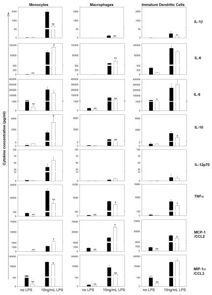
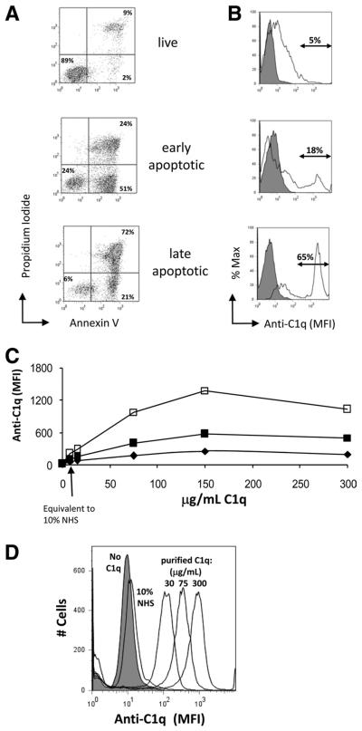
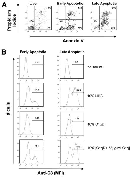
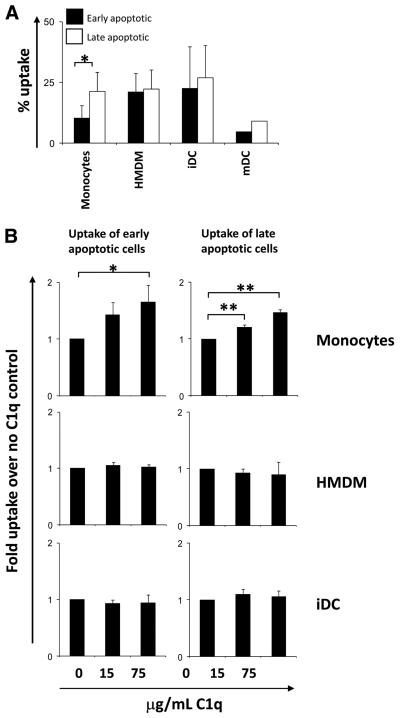
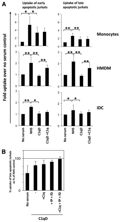
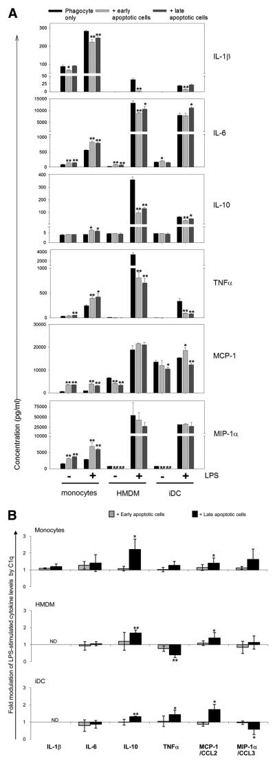
C1q, the first component of the classical complement pathway, is also a pattern recognition receptor involved in the recognition and clearance of apoptotic cells. C1q deficiency in humans leads to development of lupus-like autoimmune disease, and it has been speculated that impaired clearance of apoptotic cells may contribute to disease development. Since phagocytes initiate specific and appropriate immune responses as a result of initial ligand-receptor interactions, regulation of gene expression by C1q may also contribute to the sculpting of an immune response to the ingested "self-Ags." In this study, the role of C1q in apoptotic cell clearance and subsequent modulation of cytokine release by phagocytes was assessed including donor matched human monocytes, monocyte-derived macrophages (HMDMs), and dendritic cells (DCs). First, C1q binding is much greater to late compared with early apoptotic cells. Second, C1q binding to apoptotic cells significantly enhanced the levels of ingestion by monocytes but had no effect on HMDM and DC uptake. Third, in the presence of serum, C1q bound to apoptotic cells, activated the complement pathway, leading to C3b deposition, and enhancement of uptake of apoptotic cells by monocytes, HMDMs, and DCs. Finally, although C1q, either immobilized on a plate or bound to apoptotic cells, modulates the LPS-induced cytokine levels released by human monocytes, HMDMs, and DCs toward a more limited immune response, both the degree and direction of modulation differed significantly depending on the differentiation state of the phagocyte, providing further evidence of the integration of these cell- and environment-specific signals in determining appropriate immune responses.


Figures

FIGURE 1
FIGURE 1
Immobilized C1q differentially modulates LPS-induced cytokine release by human phagocytic cells according to their differentiation state. Cytokine levels were measured by Luminex multiplex analysis of the supernatant of phagocytic cells plated on LabTek chamber slides coated with 8 μg/ml C1q (□) or HSA control (■) and stimulated with 0 or 10 ng/ml LPS for 18 h. Data are mean values from a single experiment using donor-matched monocytes, HMDMs, and iDCs, performed in triplicate ± SD, and representative of data obtained in three experiments using different donors. *, p < 0.05; **, p < 0.005, Student's t test.
FIGURE 2
FIGURE 2
C1q binds dose-dependently to apoptotic Jurkats but not to live cells. A, Characterization of apoptotic cells. Apoptosis of Jurkat cells was induced in complete media (early apoptotic) or Xvivo serum-free media (late apoptotic). Cells were stained with Annexin V and PI and analyzed by flow cytometry. Data are from one experiment, representative of at least 10. B, Live, early apoptotic, or late apoptotic Jurkat cells were incubated with 0 μg/ml (filled histogram) or 75 μg/ml (open histogram) C1q. C, Cultures of live (◆), early apoptotic (■), or late apoptotic (□) Jurkat cells were incubated with 0–300 μg/ml human C1q for 30 min at 37°C before being washed. C1q binding was measured by the MFI of fluorescently labeled secondary Ab detection of anti-human C1q using flow cytometry. D, A typical dose-response histogram of C1q binding to late apoptotic Jurkats when incubated with purified C1q or 10% NHS. Data (B–D) are from a single experiment, representative of three.
FIGURE 3
FIGURE 3
C3b deposition on apoptotic cells. Early and late apoptotic Jurkats, as assessed by Annexin V/PI staining (A), were incubated with no serum, 10% NHS, 10% C1q-depleted serum (C1qD) (generated from the same NHS), or 10% [C1qD + 75 μg/ml C1q] for 30 min at 37°C and subsequently washed and probed with anti-C3 as described in Materials and Methods. C3 deposition was assessed by flow cytometry. Data shown are a typical histogram depicting the MFI from a single experiment, representative of three (B).
FIGURE 4
FIGURE 4
Uptake of early and late apoptotic Jurkats by monocytes, HMDMs, and iDCs. Jurkat cells were labeled with CFSE and treated with 40 μM etoposide for 16 h to induce apoptosis in complete media (early apoptotic) or serum-free Xvivo (late apoptotic). Apoptotic cells were fed to phagocytes at a ratio of 3:1 (Jurkat: monocyte/HMDM) or 1:1 (Jurkat:DC) for 30 min (HMDMs) or 60 min (monocytes and iDCs) at 37°C and the basal percent uptake determined by flow cytometry (A). Data are the mean ± SD. *, p < 0.05, Student's t test (n = 3). B, Uptake of early and late apoptotic cells was assayed in the presence or absence of C1q as indicated. Uptake was expressed as fold enhancement of percent apoptotic cell uptake over control levels (no C1q). Data are the mean ± SD. *, p < 0.05; **, p < 0.005, ANOVA, (n > 3).
FIGURE 5
FIGURE 5
Serum enhances uptake of early and late apoptotic cells. A, Early or late apoptotic Jurkat cells labeled with CFSE were incubated with phagocytes at a ratio of 3:1 (Jurkat:monocyte/HMDM) or 1:1 (Jurkat:DC) for 30 min (HMDMs) or 60 min (monocytes and iDCs) at 37°C in the absence or presence of 10% NHS, 10% C1q-depleted serum (C1qD) (generated from the same NHS), or 10% [C1qD + 140 μg/ml C1q] (C1qD+C1q). Uptake was assessed by flow cytometry and plotted as the fold uptake over no serum control. Data are the mean ± SD. *, p < 0.05; **, p < 0.005, ANOVA (n = 3, monocytes and iDCs; n = 5, HMDMs). B, Late apoptotic Jurkat cells (prelabeled with CFSE) were incubated in the absence or presence of 10% NHS, 10% C1qD, 10% [C1qD + 140 μg/ml C1q] (C1qD+C1q), 10% [C1qD + 10 μg/ml fP + 4 μg/ml fD] (C1qD +fP +fD), or 10% [C1qD + 140 μg/ml C1q + 10 μg/ml fP + 4 μg/ml fD] (C1qD +C1q +fP +fD) as indicated. Data are expressed as the mean percent uptake relative to that in the presence of 10% matched NHS of two experiments ± SD.
FIGURE 6
FIGURE 6
C1q modulation of cytokines released from phagocytes during the uptake of apoptotic cells. A, Cytokine levels were measured by Luminex multiplex analysis of the supernatant of monocytes, HMDMs, or iDCs from the same donor, plated on LabTek chamber slides, and fed early or late apoptotic cells as described in Materials and Methods and stimulated with 0 or 30 ng/ml LPS for 18 h. Data are the average concentrations ± SD from measurements of triplicate wells from a single experiment, representative of three. B, Cytokine levels were measured in the supernatant of monocytes, HMDMs, or iDCs, which had been incubated with early or late apoptotic cells opsonized with 300 μg/ml C1q for 30 min and subsequently stimulated with 30 ng/ml LPS for 18 h. Results are expressed as fold difference in expression compared with control levels from LPS-stimulated phagocytes, which had ingested apoptotic cells in the absence of C1q. Data are plotted as average fold differences ± SD from three experiments. *, p < 0.05; **, p < 0.005, C1q-treated levels vs control levels in the absence of C1q, Student's t test.
FIGURE 7
FIGURE 7
Cultured DCs secrete C1q. Secreted levels of C1q were measured by ELISA analysis of the supernatant of iDCs and mDCs plated on LabTek chamber slides coated with 8 μg/ml C1q (□) or HSA (■) control and stimulated with 0–100 ng/ml LPS for 18 h. Results are average concentration ± SD of three separate experiments. *, p < 0.05, ANOVA.

References

    1. Hart SP, Dransfield I, Rossi AG. Phagocytosis of apoptotic cells. Methods. 2008;44:280–285. - PubMed
    1. Erwig LP, Henson PM. Immunological consequences of apoptotic cell phagocytosis. Am. J. Pathol. 2007;171:2–8. - PMC - PubMed
    1. Walport MJ. Complement: second of two parts. N. Engl. J. Med. 2001;344:1140–1144. - PubMed
    1. Walport MJ. Complement: first of two parts. N. Engl. J. Med. 2001;344:1058–1066. - PubMed
    1. Bohlson SS, Fraser DA, Tenner AJ. Complement proteins C1q and MBL are pattern recognition molecules that signal immediate and long-term protective immune functions. Mol. Immunol. 2007;44:33–43. - PubMed

Publication types

MeSH terms