. 2010 Jan 8;285(2):1457-67.
doi: 10.1074/jbc.M109.038026. Epub 2009 Oct 28.

In vivo stoichiometry of shelterin components

Affiliations

In vivo stoichiometry of shelterin components

Kaori K Takai et al. J Biol Chem. .

Abstract

Human telomeres bind shelterin, the six-subunit protein complex that protects chromosome ends from the DNA damage response and regulates telomere length maintenance by telomerase. We used quantitative immunoblotting to determine the abundance and stoichiometry of the shelterin proteins in the chromatin-bound protein fraction of human cells. The abundance of shelterin components was similar in primary and transformed cells and was not correlated with telomere length. The duplex telomeric DNA binding factors in shelterin, TRF1 and TRF2, were sufficiently abundant to cover all telomeric DNA in cells with short telomeres. The TPP1.POT1 heterodimer was present 50-100 copies/telomere, which is in excess of its single-stranded telomeric DNA binding sites, indicating that some of the TPP1.POT1 in shelterin is not associated with the single-stranded telomeric DNA. TRF2 and Rap1 were present at 1:1 stoichiometry as were TPP1 and POT1. The abundance of TIN2 was sufficient to allow each TRF1 and TRF2 to bind to TIN2. Remarkably, TPP1 and POT1 were approximately 10-fold less abundant than their TIN2 partner in shelterin, raising the question of what limits the accumulation of TPP1 x POT1 at telomeres. Finally, we report that a 10-fold reduction in TRF2 affects the regulation of telomere length but not the protection of telomeres in tumor cell lines.


Figures

FIGURE 1.
FIGURE 1.
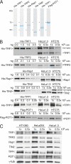
Method for quantification of TRF2. A, shown is quantification of the baculovirus (Bac)-derived His-TRF2 standard. BSA, bovine serum albumin. B, shown is quantitative immunoblotting for TRF2 in whole cell lysates of the indicated cell lines using calibration with the baculovirus-derived His-TRF2 as a standard. C, shown is the calculated abundance of TRF2 in the cell lines analyzed in B.
FIGURE 2.
FIGURE 2.
Quantitative analysis of the six shelterin components. A, shown are baculovirus-derived shelterin components used to calibrate the immunoblots. B, shown is quantitative immunoblotting for each of the shelterin components in whole cell lysates of the indicated cell lines using calibration with the baculovirus-derived proteins in A as a standard. The concentrations of the baculovirus-derived proteins were determined as in Fig. 1A. Antibodies used were: TRF1, 371; TRF2, 647; Rap1, 765; TIN2, 864; TPP1, 1150; POT1, 978. Immunoblotting for POT1 was done using guanidine renaturation. C, comparison of the abundance of shelterin components in two additional cell lines with short telomeres (HT1080 and HeLa204) to HeLa1.3.
FIGURE 3.
FIGURE 3.
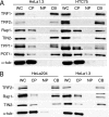
Determination of the chromatin-bound and soluble fractions of the shelterin components. A and B, the indicated cell lines were fractionated as described under “Experimental Procedures,” and equal fractions of the whole cell lysate (WC), cytoplasmic proteins (CP), nucleoplasmic proteins (NP), and the insoluble nuclear fraction, referred to as chromatin-bound proteins (CB), were analyzed by immunoblotting. α-Tubulin (α-tub) was used as a control for cytoplasmic proteins. Antibodies are as in Fig. 2.
FIGURE 4.
FIGURE 4.
Effect of shRNA-mediated knockdown of TRF2 on other shelterin components. A, shown is quantitative immunoblotting to determine the reduction of TRF2 expression in cells (HTC75) infected with retroviruses carrying shRNAs 1 and 5 compared with the luciferase (Luc) shRNA control. B, IF for TRF2 (red) at telomeres detected by fluorescence in situ hybridization (green) in HeLa1.3 cells expressing the luciferase control or sh5 to TRF2 is shown. DAPI, 4,6-diamino-2-phenylindole, C, levels of all shelterin components in whole cell lysates of the indicated cells infected with sh5 to TRF2 or the luciferase control (Lu) are shown. γ-Tubulin (γ-tub) serves as the loading control. D, shown is the effect of TRF2 knockdown on soluble and chromatin-bound shelterin components. HTC75 cells infected with the luciferase control of sh5 were fractionated as described in Fig. 3, and equal cell equivalents of the fractions were analyzed by immunoblotting for the presence of shelterin components. WC, whole cell lysate; CP, cytoplasmic proteins; NP, nucleoplasmic proteins; CB, chromatin-bound proteins.
FIGURE 5.
FIGURE 5.
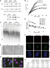
Knockdown of TRF2 results in premature senescence in primary fibroblasts. A, immunoblots of TRF2 levels in the indicated primary fibroblasts infected with TRF2 sh5 or the luciferase (Luc) controls are shown. TRF2 levels were analyzed at the indicated PDs. B, shown are growth curves of the cells described in A. PD 0 is set at day 10 after infection. C, shown is a genomic blot of telomeric restriction fragments in the cells described in A at the indicated PDs. Mr values are indicated in kb. D, shown is a graph displaying the length of the telomeric restriction fragments as a function of PD for the cells described in A. E, photographs of MRC5 expressing the indicated shRNAs at day 46 in B are shown. Cells were stained for senescence-associated β-galactosidase activity. F, shown is IF for 53BP1 (red) combined with TRF1 (green) on WI38 cells at day 10 after infection with the indicated shRNAs. The bottom images show a merge of the top two images with 4,6-diamino-2-phenylindole stain for DNA. G, example of large, non-telomeric 53BP1 foci induced by TRF2sh5 in MRC5 cells (day 7). H, a table enumerating the occurrence of TIFs and large, non-telomeric 53BP1 foci in MRC5 and HeLa1.3 treated with the indicated shRNAs is shown.
FIGURE 6.
FIGURE 6.
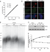
Knockdown of TRF2 affects telomere length homeostasis but not telomere protection. A, shown is a graph representing proliferation of HTC75 cells expressing the indicated shRNAs to TRF2 (sh1 and 5) or the luciferase (Luc) control. B, the absence of overt TIF phenotype in HeLa1.3 is shown. C, shown are immunoblots for TRF2 levels in whole cell lysates of HTC75 cells expressing the indicated shRNAs at early and late PD. γ-tub, γ-tubulin. D, shown is a genomic blot for telomeric restriction fragments in shRNA-treated HTC75 cells at the indicated PD. Mr markers are in kb. E, graphic representation of a second independent experiment is shown in which telomere length changes were recorded in HTC75 cells treated with sh1 or sh5 to TRF2. Methods were as D.

References

    1. Greider C. W., Blackburn E. H. (1985) Cell 43, 405–413 - PubMed
    1. Palm W., de Lange T. (2008) Annu. Rev. Genet. 42, 301–334 - PubMed
    1. Chong L., van Steensel B., Broccoli D., Erdjument-Bromage H., Hanish J., Tempst P., de Lange T. (1995) Science 270, 1663–1667 - PubMed
    1. Bilaud T., Brun C., Ancelin K., Koering C. E., Laroche T., Gilson E. (1997) Nat. Genet. 17, 236–239 - PubMed
    1. Broccoli D., Smogorzewska A., Chong L., de Lange T. (1997) Nat. Genet. 17, 231–235 - PubMed

Publication types

MeSH terms