. 2009 Nov 12;462(7270):222-5.
doi: 10.1038/nature08562. Epub 2009 Oct 28.

Human DAZL, DAZ and BOULE genes modulate primordial germ-cell and haploid gamete formation

Affiliations

Human DAZL, DAZ and BOULE genes modulate primordial germ-cell and haploid gamete formation

Kehkooi Kee et al. Nature. .

Abstract

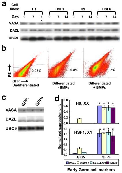
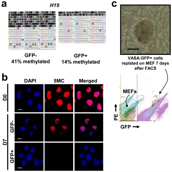
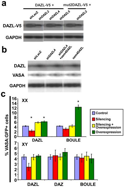
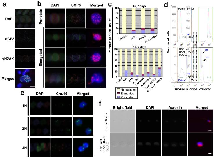
The leading cause of infertility in men and women is quantitative and qualitative defects in human germ-cell (oocyte and sperm) development. Yet, it has not been possible to examine the unique developmental genetics of human germ-cell formation and differentiation owing to inaccessibility of germ cells during fetal development. Although several studies have shown that germ cells can be differentiated from mouse and human embryonic stem cells, human germ cells differentiated in these studies generally did not develop beyond the earliest stages. Here we used a germ-cell reporter to quantify and isolate primordial germ cells derived from both male and female human embryonic stem cells. By silencing and overexpressing genes that encode germ-cell-specific cytoplasmic RNA-binding proteins (not transcription factors), we modulated human germ-cell formation and developmental progression. We observed that human DAZL (deleted in azoospermia-like) functions in primordial germ-cell formation, whereas closely related genes DAZ and BOULE (also called BOLL) promote later stages of meiosis and development of haploid gametes. These results are significant to the generation of gametes for future basic science and potential clinical applications.


Figures

Figure 1
Figure 1. Enrichment of human germ cells by BMPs and VASA:GFP reporter
a, Western analysis of hESCs after differentiation with BMPs at 7 and 14 days. Equal amounts of cell lysates were loaded in each lane. UBC9 was used as a loading control. b, FACS analysis of GFP populations. c, Western analysis of VASA and DAZL after FACS. d, Expression of early germ cell markers via qPCR Taqman probes. 20,000 GFP+ and GFP- cells (Day 7 of differentiation with BMPs) were subjected to qPCR analysis. Error bar = standard deviation; asterisk = significant difference by t-test (p<0.05), n=2.
Figure 2
Figure 2. Germ cell properties of VASA:GFP+ cells
a, GFP+ population was hypomethylated at H19 locus. b, 5-methyl cytosine staining of the VASA:GFP+ population to detect global methylation. Cells were immunostained using monoclonal 5-methyl cytosine antibody. Images are taken at the same exposure time to show different levels of staining. c, Phase contrast pictures showing representative colony from GFP+ cells after 7 days of replating. No colonies were observed from plating of GFP- population. FACS plot demonstrates that GFP+ cells maintained GFP expression after 7 day of replating on MEFs. Scale bar = 100 micron in c.
Figure 3
Figure 3. Silencing of DAZ family members and germ cell numbers
a, Western analysis of DAZL-V5 silenced by independent shDAZL constructs. DAZL with V5 epitope was cotransfected with control (shLacZ) or shDAZLs in 293T cells. Mut2DAZL-V5 was resistant to shDAZL2. b, Western analysis of DAZL and VASA after silencing with shDAZL4 or rescue in hESCs (H9). c, FACS results using H9 (XX line) and HSF1 (XY line) for silencing of DAZL, BOULE, and DAZ. Error bars = standard deviation; asterisk = significant difference in percentage of VASA:GFP+ cells by one way ANOVA, (p<0.05), n=2 (Averages from two independently differentiated samples at 14 days).
Figure 4
Figure 4. Overexpression of DAZL, DAZ and BOULE induces meiotic progression and haploid formation
a, Meiotic spread from Day 7 differentiated cells; immunofluorescence staining with SCP3 and γH2AX. b, Meiotic cells overexpressing DAZ family proteins and stained for SCP3. c, Percentage of cells showing punctate or elongated SCP3 staining at Day 7. 200 meiotic spreads were counted and categorized for each sample. Scale = 10 micron. d, FACS of DNA content of human semen and cells overexpressing DAZL, DAZ, and BOULE. e, Fluorescent in situ hybridization of chromosome 16 in cells sorted as 1N, 2N, 4N. f, ACROSIN staining of 1N population from human semen and HSF1 with 3 overexpression vectors. All cells were from whole cultures without GFP FACS.

References

    1. Hubner K, et al. Derivation of oocytes from mouse embryonic stem cells. Science. 2003;300:1251–1256. - PubMed
    1. Toyooka Y, Tsunekawa N, Akasu R, Noce T. Embryonic stem cells can form germ cells in vitro. Proc. Natl. Acad. Sci. 2003;100:11457–11462. - PMC - PubMed
    1. Geijsen N, et al. Derivation of embryonic germ cells and male gametes from embryonic stem cells. Nature. 2004;427:148–54. - PubMed
    1. Clark AT, et al. Spontaneous differentiation of germ cells from human embryonic stem cells in vitro. Hum. Mol. Genet. 2004;13:727–39. - PubMed
    1. Kee K, Gonsalves JM, Clark AT, Reijo Pera RA. Bone morphogenetic proteins induce germ cell differentiation from human embryonic stem cells. Stem Cells Dev. 2006;15:831–837. - PubMed

Publication types

MeSH terms