. 2009 Oct 29;461(7268):1300-4.
doi: 10.1038/nature08508.

Exploitation of binding energy for catalysis and design

Affiliations

Exploitation of binding energy for catalysis and design

Summer B Thyme et al. Nature. .

Abstract

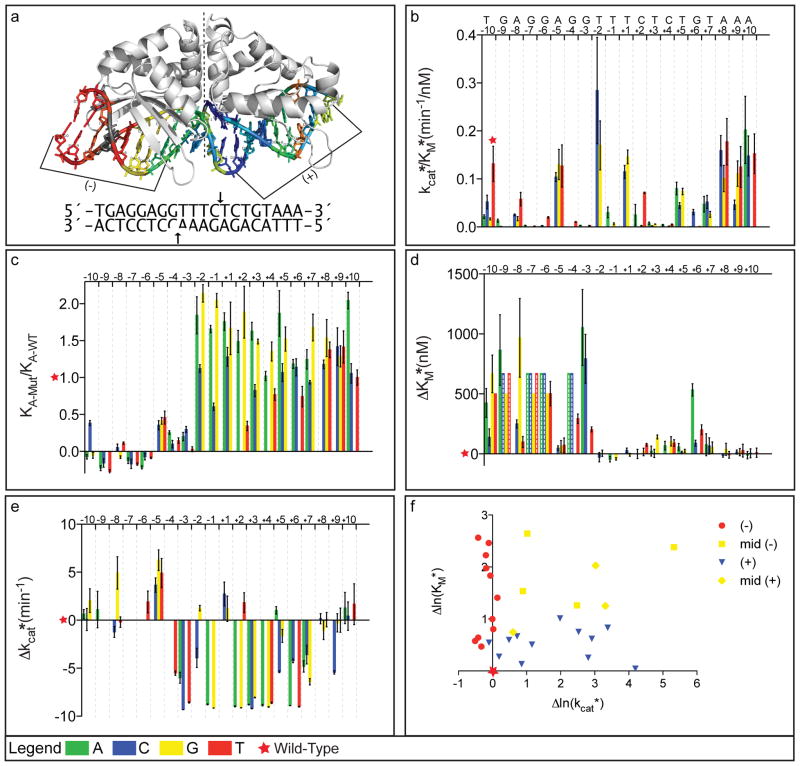
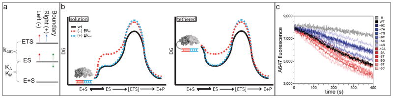
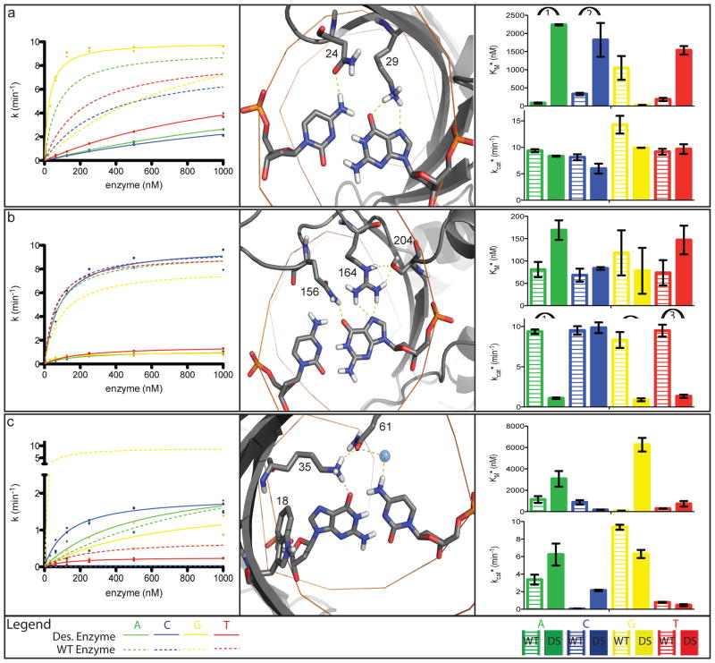
Enzymes use substrate-binding energy both to promote ground-state association and to stabilize the reaction transition state selectively. The monomeric homing endonuclease I-AniI cleaves with high sequence specificity in the centre of a 20-base-pair (bp) DNA target site, with the amino (N)-terminal domain of the enzyme making extensive binding interactions with the left (-) side of the target site and the similarly structured carboxy (C)-terminal domain interacting with the right (+) side. Here we show that, despite the approximate twofold symmetry of the enzyme-DNA complex, there is almost complete segregation of interactions responsible for substrate binding to the (-) side of the interface and interactions responsible for transition-state stabilization to the (+) side. Although single base-pair substitutions throughout the entire DNA target site reduce catalytic efficiency, mutations in the (-) DNA half-site almost exclusively increase the dissociation constant (K(D)) and the Michaelis constant under single-turnover conditions (K(M)*), and those in the (+) half-site primarily decrease the turnover number (k(cat)*). The reduction of activity produced by mutations on the (-) side, but not mutations on the (+) side, can be suppressed by tethering the substrate to the endonuclease displayed on the surface of yeast. This dramatic asymmetry in the use of enzyme-substrate binding energy for catalysis has direct relevance to the redesign of endonucleases to cleave genomic target sites for gene therapy and other applications. Computationally redesigned enzymes that achieve new specificities on the (-) side do so by modulating K(M)*, whereas redesigns with altered specificities on the (+) side modulate k(cat)*. Our results illustrate how classical enzymology and modern protein design can each inform the other.


Figures

Figure 1
Figure 1. Segregation of contributions to binding and catalysis
The color scheme throughout the figure is A=green, C=blue, G=yellow, T=red, and error bars in all panels are standard errors from the mean (SEM). a. Ribbon diagram of the I-AniI enzyme in complex with the wild-type target site (2QOJxi). Target site and positions of DNA cleavage are shown below: (−) side cleavage site is cut prior to (+) side site. b. kcat*/KM* values for the wild-type target site (red star) and each of the 60 singly-substituted target sites (vertical bars). Substitutions throughout the length of the target site abrogate enzyme activity demonstrating the high sequence specificity of the enzyme. c. Relative binding affinities determined for each singly-substituted target site using fluorescence competition assays. Substitutions on the left side, but not the right side, significantly reduce binding affinity. d. KM* values for each singly-substituted target site relative to the wild-type. As in c, substitutions on the left but not the right display significantly different values from wild-type. e. kcat* values for each singly-substituted target site relative to the wild-type site. In contrast to c and d, substitutions between positions −4 and +9 have significant effects. Substitutions for which KM* was too high (> 750nM) to allow separate determination of kcat* and KM* are indicated by bars with dashed lines in d, and are left blank in e. f. Asymmetry of the contributions to kcat* and KM*. Positions shown in red are on the left (−) side of the target site from −10 to −5 and almost exclusively contribute to KM*. Positions shown in blue are on the right (+) side of the target site from positions +3 to +7. The boundary positions, −4, −3, and +6, contribute to both kcat* and KM* and are shown in yellow. To portray the structural context of these positions, the target site in a) is colored based on the effect of the mutation on kcat*, normalized by the sum of the effects on kcat* and KM* ([|Δln(kcat*)|/|(Δln(KM*)|+ |Δln(kcat*)|)] close to 1.0, blue; close to 0.0, red; intermediate, yellow; position where KM* and kcat* could not be separately determined; grey).
Figure 2
Figure 2. Contributions to catalysis
a. Free energy diagram showing the effect of target site substitutions on the free energies of substrate binding and transition state stabilization. The majority of substitutions on the left side increase the energy of both ES and ETS, suggesting they disrupt interactions made in both states. The majority of the positions on the right side instead raise ETS, suggesting they remove interactions present in ETS but not ES. A small subset of positions (labeled boundary) appears to selectively stabilize or destabilize ES while not affecting ETS; these substitutions may disrupt interactions present in ES but not in ETS. b. Free energy profiles for a free (left) and tethered (right) system. Red profile: substitutions that remove interactions present in both ES and ETS; blue line, substitutions that remove interactions present in ETS but not ES. Tethering increases the free energy of free E + S to the point that the rate depends only on the free energy difference between ES and ETS. Since this free energy difference is unchanged by substitutions that remove interactions made in both ES and ETS (red profile), they do not affect the rate in the tethered case. c. Yeast on-cell cleavage assays. Surface displayed enzyme cleaves a tethered fluorescently labeled oligo, which then diffuses away from the yeast surface resulting in loss of fluorescence. Black, wild-type target site; random DNA, grey; shades of red, left side target site substitutions; shades of blue, right side target site substitutions. Tethering suppresses decreases in cleavage rate produced by (−) side but not (+) side mutations.
Figure 3
Figure 3. Computational redesign of specificity
The color scheme throughout the figure is A=green, C=blue, G=yellow, T=red, and error bars in right panels are standard errors from the mean (SEM). a. Design for −8A:T to −8G:C substitution (K24N, T29K). (Middle panel) The designed residues N24 and K29 make direct hydrogen bonds to −8G and −8C respectively. (Left panel) The concentration dependence of the cleavage activity for the designed enzyme (solid lines) for different base-pairs at the −8 position differs considerably from the wild type enzyme (dashed lines). (Right panel) The kcat* values remain approximately the same for both the wild-type and designed enzymes against all target sites, but the KM* values are decreased for the target G base-pair (arrow 3) and increased significantly for the other three substitutions (arrows 1, 2, and 4). b. Design for +8A:T to +8C:G substitution (L156Q, I164R, T204S). (Middle panel) Designed residues R164 and Q156 make direct hydrogen bonds to +8G. Designed residue S204 holds R164 in position. The kinetic traces (left panel) and bar graphs (right panel) show this design achieves altered specificity through changing kcat*. The KM* values remain approximately the same for both the wild-type and designed enzymes against all target sites, but the kcat* values are significantly decreased for all of the competitor target sites (arrows 1, 2, and 3). c. Design for −3G:C to −3C:G substitution (Y18W, E35K, R61Q). (Middle panel) Designed residues K35 and Q61 make a direct hydrogen bond to −3G and a water-mediated hydrogen bond to −3C, respectively. Q61 and K35 also hydrogen bond with each other, and designed residue W18 further helps position K35 through packing interactions. The kinetic traces (left panel) and bar graphs (right panel) show this design achieves altered specificity through changing both kcat* and KM*. The designed enzyme has an increased kcat* (arrow 2) and decreased KM* for the −3C (arrow 1).

References

    1. Jencks WP. Mechanism of enzyme action. Annu Rev Biochem. 1963;32:639–676. - PubMed
    1. Bolduc JM, et al. Structural and biochemical analyses of DNA and RNA binding by a bifunctional homing endonuclease and group I intron splicing factor. Genes Dev. 2003;17:2875–2888. - PMC - PubMed
    1. Wells TN, Fersht AR. Use of binding energy in catalysis measured by mutagenesis of tyrosyl-tRNA synthetase. Biochemistry. 1986;25:1881–1886. - PubMed
    1. Fersht AR. Relationships between apparent binding energies measured in site-directed mutagenesis experiments and energetics of binding and catalysis. Biochemistry. 1988;27:1577–1580. - PubMed
    1. Benkovic SJ, Hammes-Schiffer S. A perspective on enzyme catalysis. Science. 2003;301:1196–1202. - PubMed

Publication types