. 2010 Mar;17(3):424-31.
doi: 10.1038/gt.2009.144. Epub 2009 Oct 29.

Extending the transposable payload limit of Sleeping Beauty (SB) using the Herpes Simplex Virus (HSV)/SB amplicon-vector platform

Affiliations

Extending the transposable payload limit of Sleeping Beauty (SB) using the Herpes Simplex Virus (HSV)/SB amplicon-vector platform

S de Silva et al. Gene Ther. 2010 Mar.

Abstract

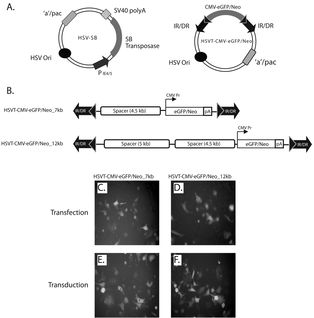
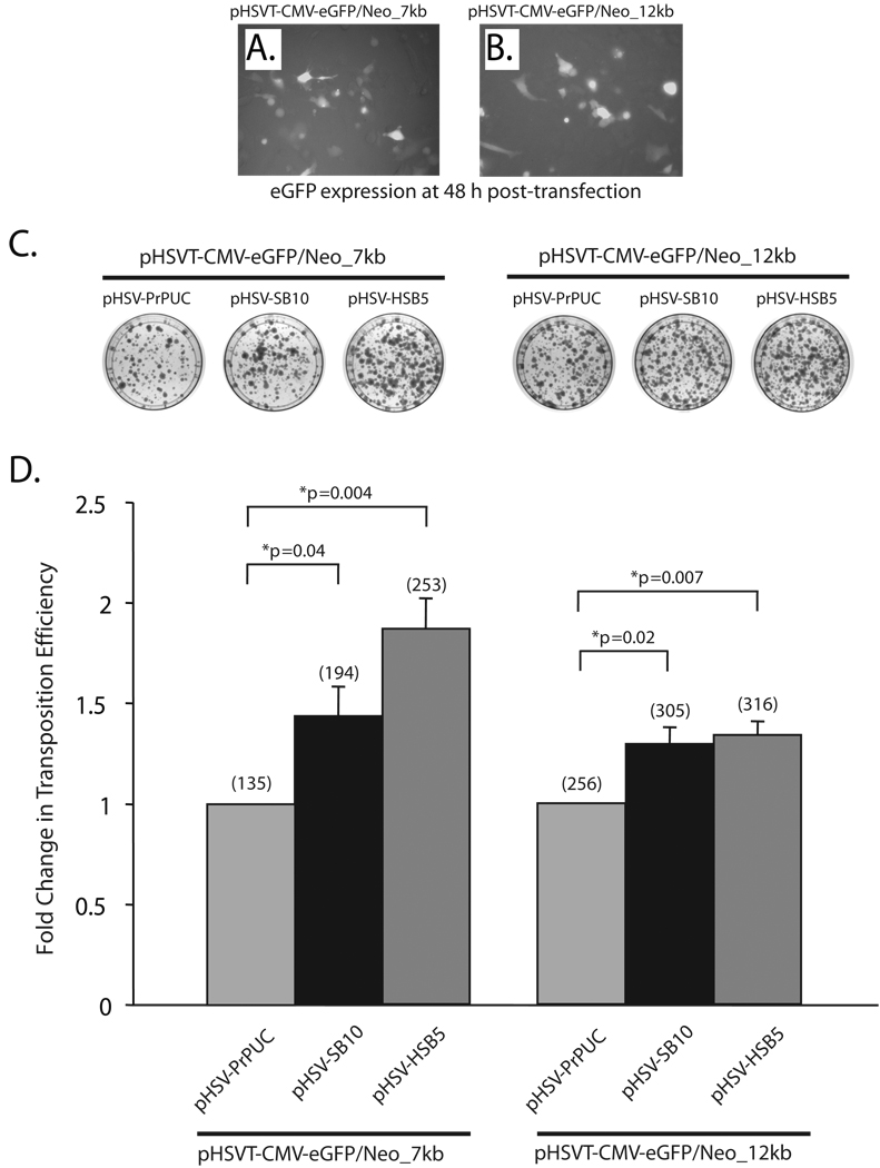
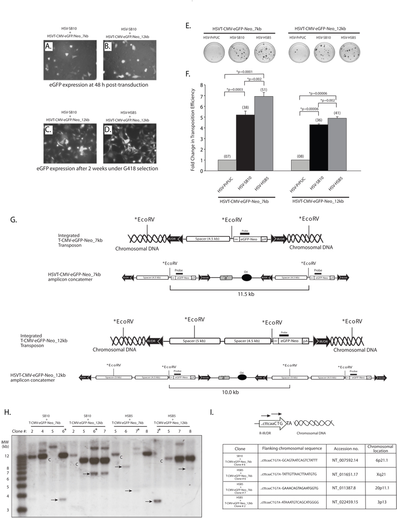
The ability of a viral vector to safely deliver and stably integrate large transgene units (transgenons), which not only include one or several therapeutic genes, but also requisite native transcriptional regulatory elements, would be of significant benefit for diseases presently refractory to available technologies. The herpes simplex virus type-1 (HSV-1) amplicon vector has the largest known payload capacity of approximately 130 kb, but its episomal maintenance within the transduced cell nucleus and induction of host cell silencing mechanisms limits the duration of the delivered therapeutic gene(s). Our laboratory developed an integration-competent version of the HSV-1 amplicon by adaptation of the Sleeping Beauty (SB) transposon system, which significantly extends transgene expression in vivo. The maximum size limit of the amplicon-vectored transposable element remains unknown, but previously published plasmid-centric studies have established that DNA segments longer than 6-kb are inefficiently transposed. Here, we compared the transposition efficiency of SB transposase in the context of both the HSV amplicon vector as well as the HSV amplicon plasmid harboring 7 and 12-kb transposable reporter transgene units. Our results indicate that the transposition efficiency of the 12-kb transposable unit via SB transposase was significantly reduced as compared with the 7-kb transposable unit when the plasmid version of the HSV amplicon was used. However, the packaged HSV amplicon vector form provided a more amenable platform from which the 12-kb transposable unit was mobilized at efficiency similar to that of the 7-kb transposable unit via the SB transposase. Overall, our results indicate that SB is competent in stably integrating transgenon units of at least 12 kb in size within the human genome upon delivery of the platform via HSV amplicons.


Figures

Figure 1
Figure 1. Schematic representation of the bipartite HSV/Sleeping Beauty amplicon vector platform and expression testing of the 7- and 12-kb transposable reporter transcription units within the plasmid and viral forms of the HSVT amplicon vector
(A) The two-component HSV/Sleeping Beauty (SB) amplicon vector platform consists of a HSV-SB amplicon vector, which expresses the SB transposase under the transcriptional regulation of the HSV IE 4/5 promoter and an integration-competent reporter amplicon vector, termed HSVT-CMV-eGFP/Neo. The latter harbors the IR/DR DNA elements of SB, which flank an enhanced green fluorescent protein gene fused to a neomycin phosphotransferase gene (eGFP-Neo) under the transcriptional regulation of the cytomegalovirus (CMV) promoter. (B) To generate 7-kb and 12-kb transposable segments, the CMV-eGFP/Neo-SV40 polyA (2.5 kb) transcription unit was excised from the pBSFBR-CMV-eGFP/Neo construct using NotI (New England BioLabs, MA) and cloned into the corresponding NotI site in a previously engineered PmeI site-containing pBSIIKS(+) shuttle vector to generate pBSIIKS/P_CMV-eGFP/Neo. Subsequently, a 4.5-kb non-coding spacer DNA fragment was obtained by digestion of pDelta28E4 (kindly provided by Dr. Brendan Lee) with Hind III, and was cloned into the corresponding Hind III site 5’ to the CMV-eGFP/Neo transcription unit to generate pBSIIKS_CMV-eGFP/Neo_4.5kb. Thereafter, the CMV-eGFP/Neo (2.5-kb) transcription unit plus the 4.5-kb spacer DNA fragment were excised using PmeI. This 7-kb segment was cloned into a blunted NotI site in the pHSVTmcs amplicon to generate the pHSVT-CMV-eGFP/Neo_7kb amplicon plasmid. To extend the size of the transposable segment to 12-kb, a 5-kb non-coding spacer fragment, which was also obtained from the pDelta28E4 plasmid via digestion with FspI, was cloned into a blunted ClaI site located upstream of the 4.5 kb-spacer fragment within pHSVT-CMV-eGFP/Neo_7kb to generate pHSVT-CMV-eGFP/Neo_12kb. Both constructs were verified by DNA sequence analysis and subsequently packaged into helper virus-free HSV amplicon particles and viral titers were determined as described previously. (C, D) To verify eGFP expression from both pHSVT-CMV-eGFP/Neo_7kb and pHSVT-CMV-eGFP/Neo_12kb amplicon plasmids, separate cultures of 5 × 10⁴ HeLa cells were transfected with each construct using FuGENE6® transfection reagent (Roche, Germany) and visualized by fluorescent microscopy 48 hrs post-transfection. (E, F) Similarly, eGFP expression derived from packaged HSV amplicon particles were confirmed by transducing HeLa cell monolayers using HSVT-CMV-eGFP/Neo_7kb and HSVT-CMV-eGFP/Neo_12kb virus at an MOI of 0.1 for 1 h at 37° C. After the 1−h incubation, virus-containing media was removed, the cells were rinsed once with media, and subsequently replenished with fresh media. eGFP expression was observed by fluorescent microscopy 48 h post-transduction.
Figure 2
Figure 2. Assessing the transposition efficiency of SB transposase in the context of HSV amplicon plasmids harboring differently sized transposable units
HeLa cells (1×10⁵ cells/well) were co-transfected with 165 ng of the plasmid versions of the HSV amplicon vectors expressing either the ‘wild-type’ SB transposase (pHSV-SB10) or the hyperactive HSB5 transposase (pHSV-HSB5), together with either 116 ng of the pHSVT-CMV-eGFP/Neo_7kb amplicon or 165 ng of the pHSVT-CMV-eGFP/Neo_12kb amplicon using the FuGENE6® transfection reagent (Roche, Germany). The pHSVPrPUC empty vector plasmid was transfected with each of the differently sized eGFP/Neo transposon-harbored amplicons to determine the number of G418-resistant colonies that arise due to random integration events in the absence of SB transposase. To ensure equal amounts of DNA were transfected under each condition, the pHSVPrPUC empty vector was used as ”filler” DNA. (A, B) eGFP expression was assessed 48 h post-transfection via fluorescence microscopy to determine the transfection efficiency of the pHSVT-CMV-eGFP/Neo_7kb and the pHSVT-CMV-eGFP/Neo_12kb amplicon plasmids in the presence of the SB-expressing amplicon plasmid. (C) Thereafter, the cells were trypsinized and seeded at a 1:3 dilution on 100-mm dishes and placed under 600 µg/ml G418 selection for a period of 2 weeks. Subsequently, the cells were fixed with 4% paraformaldehyde and stained with 2% methylene blue, washed extensively with dH2O and blue colonies were enumerated. (D) The transposition efficiency of SB10 and HSB5 in the presence of either the 7- or 12-kb transposons is represented as a fold-change in the number of G418-resistant colonies compared to when SB transposase was absent. The average number of G418-resistant colonies (n=3) corresponding to each group is indicated in parentheses above each bar in the histogram. Error bars represent standard error. Statistical analysis was conducted using the student t-test and p values are indicated.
Figure 3
Figure 3. The SB transposase is competent in efficiently mobilizing a 12-kb transposable unit from the context of an HSV amplicon vector
HeLa cells (1×10⁵ cells/well) were co-transduced with HSV amplicon vectors expressing either the “wild-type” SB transposase (HSV-SB10) or the hyperactive HSB5 transposase (HSV-HSB5), together with either the HSVT-CMV-eGFP/Neo_7kb amplicon or the HSVT-CMV-eGFP/Neo_12kb amplicon at an MOI of 1.0. Upon incubation for 1 h at 37°C, the virus-containing medium was removed and cells were washed once and replenished with fresh media. (A–B) Enhance GFP expression was assessed 48 h post-transduction via fluorescence microscopy to determine the transduction efficiency of the HSVT-CMV-eGFP/Neo_7kb and HSVT-CMV-eGFP/Neo_12kb amplicons in the presence of the co-delivered SB-expressing amplicon vector. Subsequently, the cells were trypsinized and seeded at a 1:3 dilution on 100-mm dishes and placed under 600 µg/ml G418 selection for a period of 2 weeks. (C–D) After 2 weeks, G418-resistant colonies were fixed with 4% paraformaldehyde and analyzed via fluorescence microscopy for eGFP expression. Representative images of G418-resistant colonies 2 weeks post-transduction of HSVT-CMV-eGFP/Neo_12kb either with HSV-SB10 or HSV-SB12 are shown. (E) Upon confirmation of eGFP expression, the G418-resistant colonies were stained with 2% methylene blue, washed extensively with dH2O, and blue colonies were enumerated. (F) The transposition efficiency of SB10 and HSB5 in the presence of either the 7- or 12-kb transposon-harboring HSVT amplicons is represented as a fold-change in the number of G418-resistant colonies compared to when SB transposase was absent (HSVPrPUC control amplicon). The average number of G418-resistant colonies (n=3) corresponding to each group is indicated in parentheses above each bar in the histogram. Error bars represent standard error. Statistical analysis was conducted using the student t-test and p values are indicated. (G) Schematic representations of integrated configurations of the T-CMV-eGFP-Neo_7kb and T-CMV-eGFP-Neo_12kb transgenons and their concatemeric amplicon genome counterparts. The relative locations of signature EcoRV restriction enzyme recognition sites and the location of a DNA probe used in subsequent Southern blot analyses are depicted. (H) Similarly, G418-resistant colonies that were co-transduced with either HSV-SB10 or HSV-HSB5 and HSVT-CMV-eGFP/Neo_7kb or HSVT-CMV-eGFP/Neo_12kb were trypsinized and expanded in 60-mm dishes for the purpose of isolating genomic DNA for Southern blot analysis to determine chromosomal integration of the 7- and 12-kb transposons. Once confluent, these transduced HeLa cell monolayers were lysed using lysis buffer (10 mM Tris.Cl, 100 mM NaCl, 25 mM EDTA and 0.5% SDS) and genomic DNA was isolated using phenol:chloroform extraction followed by ethanol precipitation. Ten micrograms of genomic DNA was digested with EcoRV, which cuts once within the 7-kb transposable unit 5’ of the CMV-eGFP-Neo transcription unit and twice within the 12-kb transposable unit. The digested genomic DNA was electrophoresed on a 0.8% TAE agarose gel and transferred onto a nylon membrane, UV cross-linked and probed with a α-32P-dCTP-radiolabeled eGFP/Neo probe (917-bp), which was obtained by excising the eGFP-Neo fragment from the pBSFBR-CMV-eGFP-Neo vector using Pst1 and BamHI. The 1-kb plus DNA ladder (Invitrogen, Carlsbad, CA) was radiolabeled according to manufacturer recommendations (Lane 1). “C” indicates HSV amplicon units generated by EcoRV digestion of the episomal HSV amplicon concatemer or integrated concatemeric transgenon units, while the ‘arrows’ indicate predicted SB-mediated integration events within the HeLa cell genome and “*” demarcates clones later analyzed by integration site mapping. (I) To determine the integration sites of the 7-kb and 12-kb transposable units within the HeLa cell genome, linker-mediated PCR (LM-PCR) analysis was conducted using the GenomeWalker™ Universal Kit (Clontech, Mountain view, CA) according to manufacturer recommendations. Chromosomal sequences flanking the right IR/DR junction of the 7- and 12-kb transposable units were PCR amplified from genomic DNA samples that were analyzed by Southern blotting using a nested primer set described in Largaespada et al., and cloned into the TOPO-TA vector (Invitrogen, Carlsbad, CA) for subsequent sequence analysis.

Similar articles

Cited by

References

    1. Hermonat PL, Quirk JG, Bishop BM, Han L. The packaging capacity of adeno-associated virus (AAV) and the potential for wild-type-plus AAV gene therapy vectors. FEBS Lett. 1997;407:78–84. - PubMed
    1. Kumar M, Keller B, Makalou N, Sutton RE. Systematic determination of the packaging limit of lentiviral vectors. Hum Gene Ther. 2001;12:1893–1905. - PubMed
    1. Senior SL, Wade-Martins R. Herpes simplex virus type 1 amplicon vectors for the infectious delivery and expression of genomic DNA loci. Curr Opin Mol Ther. 2005;7:337–345. - PubMed
    1. Suzuki M, Kasai K, Saeki Y. Plasmid DNA sequences present in conventional herpes simplex virus amplicon vectors cause rapid transgene silencing by forming inactive chromatin. J Virol. 2006;80:3293–3300. - PMC - PubMed
    1. Wang Y, et al. Herpes simplex virus type 1/adeno-associated virus rep(+) hybrid amplicon vector improves the stability of transgene expression in human cells by site-specific integration. J Virol. 2002;76:7150–7162. - PMC - PubMed

Publication types

Substances