Review
. 2010 Jan;67(1):99-111.
doi: 10.1007/s00018-009-0155-7. Epub 2009 Oct 29.

Melanopsin and inner retinal photoreception

Affiliations
Review

Melanopsin and inner retinal photoreception

Helena J Bailes et al. Cell Mol Life Sci. 2010 Jan.

Abstract

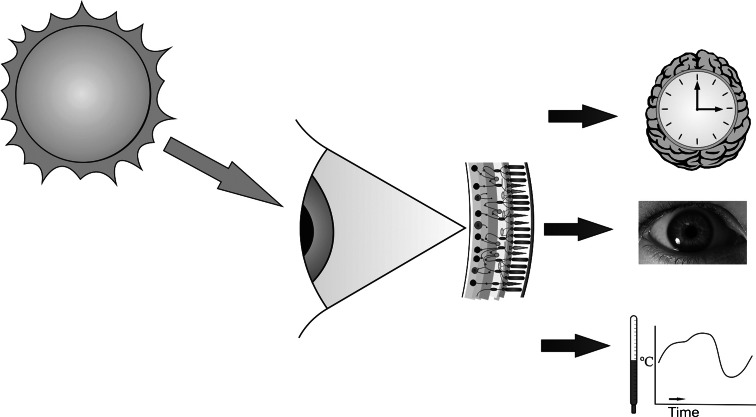
Over the last ten years there has been growing acceptance that retinal photoreception among mammals extends beyond rods and cones to include a small number of intrinsically photosensitive retinal ganglion cells (ipRGCs). These ipRGCs are capable of responding to light in the absence of rod/cone input thanks to expression of an opsin photopigment called melanopsin. They are specialised for measuring ambient levels of light (irradiance) for a wide variety of so-called non-image-forming light responses. These include synchronisation of circadian clocks to light:dark cycles and the regulation of pupil size, sleep propensity and pineal melatonin production. Here, we provide a review of some of the landmark discoveries in this fast developing field, paying particular emphasis to recent findings and key areas for future investigation.


Figures

Fig. 1
Fig. 1
A schematic figure of the non-image-forming (NIF) visual system in mammals involving ipRGCs. Light falling on the retina is detected by rod and cone photoreceptors and by the melanopsin phototransduction cascade in ipRGCs. The resultant signals are combined by ipRGCs to send an integral signal of irradiance to the brain. In turn, light information is passed to the brain down the optic nerve, triggering a diverse range of non image-forming responses in addition to normal form vision. NIF responses comprise the entrainment of circadian rhythms including sleep propensity (shown here by an image of a clock superimposed on a brain), the pupil light reflex, regulation of pineal melatonin levels and, in humans, regulation of body temperature and mood
Fig. 2
Fig. 2
A schematic diagram of retinal connections relevant to ipRGCs in the rodent retina. A and B show examples of classical wiring through ON (triggered by increments of light) and OFF (triggered by decrements of light)-cell pathways of the retina, respectively. The typical pathway of vertical transmission flows from photoreceptor cell to bipolar cell to ganglion cell. The inner plexiform layer (IPL) can be subdivided into sublaminae a and b. Bipolar and ganglion cells stratifying in sublamina a are OFF cells, while those stratifying in sublamina b are ON. A cone ON bipolar cell synapses directly onto an ON ganglion cell in sublamina b. Rod bipolar cells are all ON cells but do not contact ganglion cells directly in this classical pathway, but rather synapse onto AII amacrine cells in sublamina b. AII amacrines in turn contact cone ON bipolar cell dendrites through sign-conserving gap junctions, and OFF cone bipolar cells via sign inverting synapses (B). Cone OFF bipolar cells directly synapse onto OFF ganglion cells. Diverse amacrine cell types modulate ON and OFF pathways mainly through inhibitory inputs. ipRGCs can be subdivided into three morphological types (M1–M3). M1 cell dendrites stratify in sublamina a (C), M2 in sublamina b (B), while M3 cells are bistratified in both sublaminae (D). In contrast to normal retinal cell stratification patterns, however, M1 cells stratifying in sublamina a receive strong ON signals [43, 47, 48]. This reflects direct input from cone ON bipolars at the M1 cell body and, anomalously at dendrites in sublamina a [49, 50], as well as direct synaptic contacts with rod bipolars [47]. Rod bipolars may also influence their activity via the classical AII amacrine cell route. Physiological results predict additional input from cone OFF bipolars and inhibitory amacrine cells [51, 52]. Finally, there are direct connections between dopaminergic amacrine cells and presumed M1 cell dendrites in sublamina a, possibly going in both directions [46, 47, 50, 53, 54]. The connections of M2 and M3 cells are currently unknown. Those shown here are predictions based on connectivity of conventional ganglion cells with similar dendritic stratification patterns. Not shown are horizontal cells which modulate information transfer at the OPL. ONL outer nuclear layer, OPL outer plexiform layer, INL inner nuclear layer, IPL inner plexiform layer, GCL ganglion cell layer, HC horizontal cell, AC amacrine cell, AC* aII amacrine cell, AC+ dopaminergic amacrine cell, BC, bipolar cell. Blue bipolar cell body, cone ON bipolar; red bipolar cell body, rod bipolar (ON); grey bipolar cell body, cone OFF bipolar
Fig. 3
Fig. 3
Melanopsin is maximally sensitive to short wavelength ‘blue’ light. The spectral sensitivity profile of melanopsin photoreception in a standard human observer is approximated by the nomogram for an opsin:vitamin A based photopigment with peak sensitivity at 480 nm corrected for likely wavelength-dependent lens absorption. The spectral sensitivity of scotopic (rod-based) and photopic (cone-based) vision (Commission Internationale de l’Eclairage, CIE, luminosity functions) are presented for comparison. A very rough approximation of perceived colour is shown below. Note that although melanopsin sensitivity peaks around 480 nm, it shows good responsiveness well into the green/yellow portion of the spectrum

References

    1. Takahashi JS, DeCoursey PJ, Bauman L, Menaker M. Spectral sensitivity of a novel photoreceptive system mediating entrainment of mammalian circadian rhythms. Nature. 1984;308:186–188. - PubMed
    1. Foster RG, Helfrich-Forster C. The regulation of circadian clocks by light in fruitflies and mice. Philos Trans R Soc Lond B. 2001;356:1779–1789. - PMC - PubMed
    1. Nelson DE, Takahashi JS. Sensitivity and integration in a visual pathway for circadian entrainment in the hamster (Mesocricetus auratus) J Physiol. 1991;439:115–145. - PMC - PubMed
    1. von Frisch K. Beiträge zur Physiologie der Pigmentellen in der Fischhaut. Pflügers Arch. 1911;138:319–387.
    1. Tamotsu S, Morita Y. Photoreception in pineal organs of larval and adult lampreys, Lampetra japonica . J Comp Physiol A. 1986;159:1–5. - PubMed

Publication types

LinkOut - more resources