. 2010 Jan;19(1):34-46.
doi: 10.1002/pro.280.

Thermodynamic and structural insights into CSL-DNA complexes

Affiliations

Thermodynamic and structural insights into CSL-DNA complexes

David R Friedmann et al. Protein Sci. 2010 Jan.

Abstract

The Notch pathway is an intercellular signaling mechanism that plays important roles in cell fates decisions throughout the developing and adult organism. Extracellular complexation of Notch receptors with ligands ultimately results in changes in gene expression, which is regulated by the nuclear effector of the pathway, CSL (C-promoter binding factor 1 (CBF-1), suppressor of hairless (Su(H)), lin-12 and glp-1 (Lag-1)). CSL is a DNA binding protein that is involved in both repression and activation of transcription from genes that are responsive to Notch signaling. One well-characterized Notch target gene is hairy and enhancer of split-1 (HES-1), which is regulated by a promoter element consisting of two CSL binding sites oriented in a head-to-head arrangement. Although previous studies have identified in vivo and consensus binding sites for CSL, and crystal structures of these complexes have been determined, to date, a quantitative description of the energetics that underlie CSL-DNA binding is unknown. Here, we provide a thermodynamic and structural analysis of the interaction between CSL and the two individual sites that comprise the HES-1 promoter element. Our comprehensive studies that analyze binding as a function of temperature, salt, and pH reveal moderate, but distinct, differences in the affinities of CSL for the two HES-1 binding sites. Similarly, our structural results indicate that overall CSL binds both DNA sites in a similar manner; however, minor changes are observed in both the conformation of CSL and DNA. Taken together, our results provide a quantitative and biophysical basis for understanding how CSL interacts with DNA sites in vivo.


Figures

Figure 1
Figure 1
HES-1 SPS and CSL structure. (A) Nucleotide sequence and schematic representation of the mammalian HES-1 SPS (top) and the oligomeric DNA duplexes corresponding to the consensus and nonconsensus sites of the HES-1 SPS (bottom) that were used in the herein described binding studies. CSL-binding sites are in bold text and arrows denote directionality of binding sites. The T/A base pair that deviates from the consensus is colored red. (B) Ribbon diagram for mouse CSL-DNA structure (3BRG). The NTD, BTD, and CTD are colored cyan, green, and orange, respectively. The DNA is in a stick representation with carbon, oxygen, nitrogen, and phosphorous atoms colored grey, red, blue, and orange, respectively. The DNA from this complex structure corresponds to the HES-1 consensus binding site.
Figure 2
Figure 2
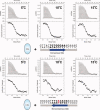
CSL-DNA ITC binding assays. Figure shows representative thermograms (raw heat signal and nonlinear least squares fit to the integrated data) for CSL binding to DNA corresponding to the HES-1 5′ consensus site (top) and the 3′ nonconsensus site (bottom). Data were measure at 5°C, 10°C, and 15°C in a phosphate buffer at pH 6.5 with 150 mM NaCl. Forty titrations were performed per experiment, consisting of 7-μl injections of DNA that were spaced 120 sec apart. [Color figure can be viewed in the online issue, which is available at www.interscience.wiley.com.]
Figure 3
Figure 3
Thermodynamic profiles for CSL binding to the consensus and nonconsensus HES-1 DNA sites. Figure shows plots of thermodynamic parameters for CSL binding to the consensus site (A) and nonconsensus site (B) that comprise the HES-1 SPS. A straight line was fit to data collected at 5°C, 10°C, and 15°C, highlighting the compensatory changes in enthalpy (ΔH°) and entropy (−TΔS°) as a function of temperature, which maintain a relatively temperature independent free energy of binding (ΔG°). The heat capacity change (ΔCp) was calculated from the slope of the line fit to the enthalpic data measured at 5°C, 10°C, and 15°C. The ΔCp values for CSL binding to the consensus and nonconsensus sites are −0.31 and −0.30 kcal/mol/K, respectively.
Figure 4
Figure 4
Salt dependence of CSL binding to DNA. Figure shows the plot of log Ka (association constant) versus the log of NaCl concentration for CSL binding to the HES-1 consensus DNA site (▪) and nonconsensus DNA site (▴). Binding experiments were performed at 50, 100, 125, 150, 175, and 200 mM NaCl.
Figure 5
Figure 5
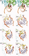
BTD-DNA interactions. Figure shows the structural details of the BTD interaction with DNA and highlights the conformational differences in a β-hairpin loop motif between structures. Wall-eye stereo pairs are depicted. (A) Simplified overview of interactions between the BTD of CSL and DNA, showing the insertion of the β-hairpin loop motif into the minor groove of DNA. The cα trace for the NTD and BTD of CSL are colored cyan and green, respectively. The DNA backbone is represented as a ribbon and colored orange. The β-hairpin loop motifs from the murine CSL consensus DNA structure (3BRG), the human ternary complex structure (2F8X), and the murine CSL nonconsensus DNA structure (3IAG) are colored green, red, and blue, respectively. Note the high degree of structural correspondence between 2F8X (red) and 3IAG (blue), but the large structural change in the β-hairpin loop motif for 3BRG (green). (B) Zoom view of the BTD interaction with DNA from the human CSL-NotchIC-Mastermind ternary complex structure (2F8X). The β-hairpin loop is in a stick representation with standard coloring for the atoms (yellow, red, and blue for carbon, oxygen, and nitrogen atoms, respectively). The DNA is also in a stick representation and colored grey, red, blue, and orange for carbon, oxygen, nitrogen, and phosphorous atoms, respectively. For clarity, only three base pairs of the DNA are shown (-TGTGGGAA-). Hydrogen bonds are indicated with black dashed lines. As also observed in previous CSL structures (e.g., 1TTU and 2FO1), Q182 makes hydrogen-bonding interactions with the adenine in the first T/A base step and the backbone carbonyl of S181 (denoted with a cyan asterisk) makes hydrogen-bonding interactions with the guanine at the following G/C base step. Nonspecific interactions with the DNA backbone mediated by residues R178, R180, and T183 are also shown, and the backbone carbonyl of R180 is indicated with a magenta asterisk. (C) Corresponding zoom view of murine CSL nonconsensus DNA complex structure determined here (3IAG). Orthologous residues and hydrogen bonding pattern are depicted, demonstrating identical interactions as described earlier. (d) Zoom view of murine CSL consensus DNA complex structure (3BRG), highlighting structural rearrangement of β-hairpin loop. In this conformation of the BTD loop, the side chains of Q222 and T223 and the backbone carbonyl of S221 no longer form interactions with the DNA; however, the rearrangement allows the side chain of S221 to make hydrogen-bonding interactions with the adenine in the T/A base step (-TGTGGGAA-) and the backbone carbonyl of R220 to make hydrogen-bonding interactions with the guanine in the G/C base step (-TGTGGGAA-). Thus, despite the rearrangement, equivalent interactions with the DNA are maintained.

References

    1. Artavanis-Tsakonas S, Rand MD, Lake RJ. Notch signaling: cell fate control and signal integration in development. Science. 1999;284:770–776. - PubMed
    1. Tanigaki K, Honjo T. Regulation of lymphocyte development by Notch signaling. Nat Immunol. 2007;8:451–456. - PubMed
    1. Gridley T. Notch signaling and inherited disease syndromes. Hum Mol Genet. 2003;12:R9–R13. - PubMed
    1. High FA, Epstein JA. The multifaceted role of Notch in cardiac development and disease. Nat Rev Genet. 2008;9:49–61. - PubMed
    1. Aster JC, Pear WS, Blacklow SC. Notch signaling in leukemia. Annu Rev Pathol. 2008;3:587–613. - PMC - PubMed

Publication types

MeSH terms

Substances

Associated data

LinkOut - more resources