. 2010 Feb;25(2):200-10.
doi: 10.1359/jbmr.090806.

Wnt inhibitors Dkk1 and Sost are downstream targets of BMP signaling through the type IA receptor (BMPRIA) in osteoblasts

Affiliations

Wnt inhibitors Dkk1 and Sost are downstream targets of BMP signaling through the type IA receptor (BMPRIA) in osteoblasts

Nobuhiro Kamiya et al. J Bone Miner Res. 2010 Feb.

Abstract

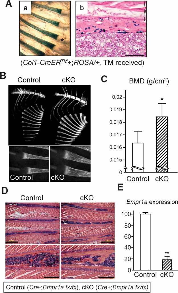
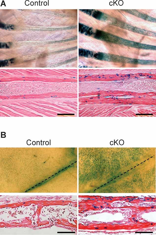
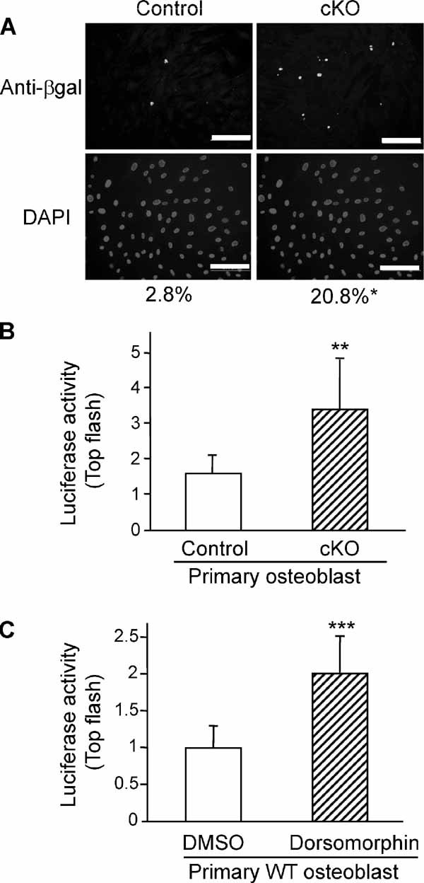
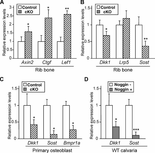
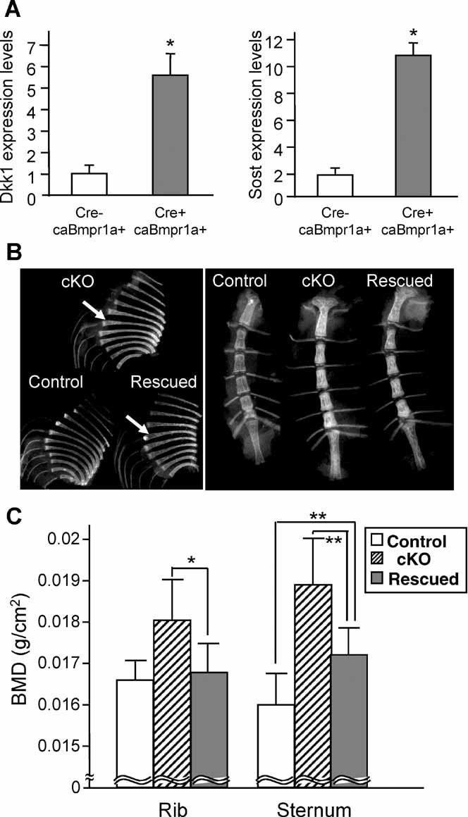
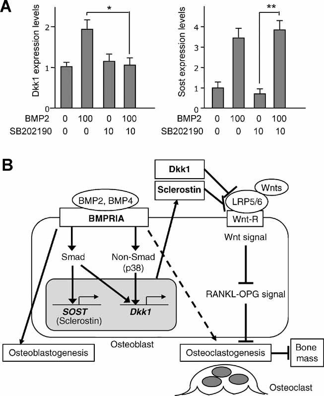
The bone morphogenetic protein (BMP) and Wnt signaling pathways both contribute essential roles in regulating bone mass. However, the molecular interactions between these pathways in osteoblasts are poorly understood. We recently reported that osteoblast-targeted conditional knockout (cKO) of BMP receptor type IA (BMPRIA) resulted in increased bone mass during embryonic development, where diminished expression of Sost as a downstream effector of BMPRIA resulted in increased Wnt/beta-catenin signaling. Here, we report that Bmpr1a cKO mice exhibit increased bone mass during weanling stages, again with evidence of enhanced Wnt/beta-catenin signaling as assessed by Wnt reporter TOPGAL mice and TOPFLASH luciferase. Consistent with negative regulation of the Wnt pathway by BMPRIA signaling, treatment of osteoblasts with dorsomorphin, an inhibitor of Smad-dependent BMP signaling, enhanced Wnt signaling. In addition to Sost, Wnt inhibitor Dkk1 also was downregulated in cKO bone. Expression levels of Dkk1and Sost were upregulated by BMP2 treatment and downregulated by Noggin. Moreover, expression of a constitutively active Bmpr1a transgene in mice resulted in the upregulation of both Dkk1 and Sost and partially rescued the Bmpr1a cKO bone phenotype. These effectors are differentially regulated by mitogen-activated protein kinase (MAPK) p38 because pretreatment of osteoblasts with SB202190 blocked BMP2-induced Dkk1 expression but not Sost. These results demonstrate that BMPRIA in osteoblasts negatively regulates endogenous bone mass and Wnt/beta-catenin signaling and that this regulation may be mediated by the activities of Sost and Dkk1. This study highlights several interactions between BMP and Wnt signaling cascades in osteoblasts that may be amenable to therapeutic intervention for the modification of bone mass density.


Figures

Fig. 1
Fig. 1
Increased bone mass in Bmpr1a cKO weanling mice. Tamoxifen was injected intraperitoneally into nursing females every 3 days from P2 to P20. (A) ROSA26 Cre reporter mice showed Cre activity on rib bones stained with β-galactosidase (β-gal) at P21 (a). Cre-dependent DNA recombination was detected in osteoblasts in rib bones by β-gal staining (b). The asterisk indicates negative staining for β-gal on rib cartilage. Bar: 100 µm. (B) Radiodensity of rib bones and sternum was increased in cKO mice (Cre+, Bmpr1afx/fx) compared with controls (Cre, Bmpr1afx/fx) when assessed by X-ray imaging at P21. Rib flaring in cKO mice is shown (right panel). (C) BMD was determined in ribs using DXA at P21. Values are expressed as mean ± SD (control: n = 10; cKO: n = 12). The asterisk indicates a statistically significant difference between control and cKO (mean ± SD; t test; *p < .04). (D) H&E staining of P21 rib bones. Rib bones were thicker in cKO mice, and bone mass was increased compared with littermate controls in both the diaphysis (upper panels) and metaphysis (lower panels). Bars: 1 mm (upper panels), 500 µm (lower panels). (E) qRT-PCR analysis for Bmpr1a using P21 rib bones (mean ± SD; t test; **p < .02).
Fig. 2
Fig. 2
Upregulation of canonical Wnt signaling in Bmpr1a cKO mice. (A) Canonical Wnt signaling in P14 rib bones assessed using TOPGAL mice. Whole-mount β-gal staining showed increased staining in cKO rib bones compared with controls (upper panels). Histologic analysis showed an increased number of β-gal-positive osteoblasts in cKO rib bones compared with controls (lower panels). Bar: 200 µm. (B) Canonical Wnt signaling in P10 skull bones assessed using TOPGAL mice. Whole-mount β-gal staining showed increased staining in cKO calvariae compared with controls (upper panels). Histologic analysis showed an increased number of β-gal-positive osteoblasts in cKO calvariae compared with controls (lower panels). Dotted lines indicate the sagittal suture. Bar: 100 µm.
Fig. 3
Fig. 3
Upregulation of canonical Wnt signaling by loss of BMP signaling in vitro. (A) Detection of canonical Wnt signaling in primary osteoblasts from P10 Bmpr1a cKO and control calvariae (upper panels) using TOPGAL mice assessed by immunocytochemical staining for β-gal (green). Nuclei were stained with DAPI (blue, lower panels). Relative ratio of β-gal-positive cell number to DAPI-positive cell number was determined in the same field (mean from 50 fields; t test; *p < .01; n > 4 per each genotype). Bars: 200 µm. (B) TOPFLASH plasmid was cotransfected with pRL-actin vector into primary osteoblasts from newborn Bmpr1a cKO and wild-type control calvariae using FuGENE 6 for 48 hours. Transfection efficiency for luciferase activity was normalized to Renilla luciferase (pRL-actin vector) activity. The y axis shows relative luciferase units (RLUs, luciferase activity/Renilla units). Promoter activities for Lef/Tcf-dependent transcription were increased significantly in cKO osteoblasts (mean ± SD; t test; **p < .02). (C) Effects of BMP signaling inhibitor dorsomorphin on canonical Wnt signaling in vitro. TOPFLASH plasmid was cotransfected with pRL-actin vector into primary osteoblasts from newborn wild-type calvariae using FuGENE 6. After 40 hours of transfection, these cells were treated with dorsomorphin (10 µM) or DMSO without serum for 8 hours, and then cells were harvested. The Lef/Tcf-dependent transcriptional activities were increased significantly by the treatment with dorsomorphin, as measured by dual luciferase activity (mean ± SD; t test; ***p < .05).
Fig. 4
Fig. 4
Downregulation of Dkk1 and Sost in Bmpr1a cKO rib bones. (A) qRT-PCR analysis for Wnt target genes Axin2, Ctgf, and Lef1 using P21 rib bones. Expression levels of Axin2, Ctgf, and Lef1 were increased significantly in cKO rib bones. Values in cKO bones (striped bar) are expressed relative to controls (open bar). (B) qRT-PCR analysis for Dkk1, Lrp5, and Sost using P21 rib bones. Expression levels of Wnt inhibitors (Dkk1 and Sost) were reduced significantly in cKO rib bones (striped bar), whereas expression of coreceptor Lrp5 was unchanged. (C) qRT-PCR analysis for Dkk1, Sost, and Bmpr1a using primary osteoblasts from P10 Bmpr1a cKO and control calvariae. Expression levels of Dkk1, Sost, and Bmpr1a were reduced significantly in cKO osteoblasts (striped bar) compared with control osteoblasts (open bar). (D) Suppressed expression of Dkk1 and Sost by Noggin treatment ex vivo, as assessed by qRT-PCR. Newborn calvariae from wild-type mice were treated with Noggin (100 ng/mL) for 5 days. The asterisk indicates a statistically significant difference between Noggin treatment (Noggin+) and nontreatment (Noggin) from three independent experiments. (A–D) Mean ± SD; t test; *p < .05; **p < .02; ***p < .01.
Fig. 5
Fig. 5
Positive regulation of Dkk1 and Sost expression by BMP2. Primary osteoblasts were isolated from wild-type newborn calvariae. (A) Dose-dependent effects of BMP2 on Dkk1 and Sost expression as assessed by qRT-PCR. mRNA was isolated from wild-type osteoblasts treated with BMP2 at the indicated concentration for 3 hours. Values are expressed relative to nontreated osteoblasts (mean ± SD; t test; *p < .05; **p < .01). (B) Time course of Dkk1 and Sost expression levels using wild-type osteoblasts treated with BMP2 (100 ng/mL). (C) Effects of pretreatments of primary osteoblasts with Smad-dependent inhibitor dorsomorphin. mRNA was isolated from wild-type osteoblasts pretreated with dorsomorphin (10 µM) or DMSO in the absence of serum for 1 hour prior to the addition of BMP2 (100 ng/mL) or PBS for 3 hours. Upregulation of Dkk1 and Sost expression by BMP2 treatment was restored by dorsomorphin pretreatment, as assessed by qRT-PCR. Values are expressed relative to osteoblasts without dorsomorphin and BMP2 (mean ± SD; t test; *p < .05).
Fig. 6
Fig. 6
Effects of Smad-dependent Bmpr1a signaling on Dkk1 and Sost expression. (A) Positive regulation of Dkk1 and Sost expression by overexpressing Smad-dependent Bmpr1a signaling. Primary osteoblasts were isolated from constitutively active Bmpr1a (caBmpr1a) newborn mice or littermate controls (Cre–, caBmpr1a+) and were treated with 4-hydroxyl tamoxifen (100 ng/mL). Both Dkk1 expression and Sost expression were increased significantly more than 5- and 10-fold, respectively, in the caBmpr1a mutant osteoblasts (Cre+, caBmpr1a+) compared with controls (Cre, caBmpr1a+), as assessed by qRT-PCR. Values of the mutant osteoblasts (striped bar) are expressed relative to controls (open bar) (mean ± SD; t test; *p < 0.01). (B) Radiodensity of rib bones and sternum from rescued mice expressing caBmpr1a on a Bmpr1a cKO background (Cre+, caBmpr1a+, Bmpr1afx/fx), littermate Bmpr1a cKO mice (Cre+, caBmpr1a, Bmpr1afx/fx) and littermate controls (Cre, caBmpr1a+, or Bmpr1afx/fx) at P21 when assessed by X-ray imaging. White arrows indicate rib flaring observed in cKO and rescued mice. (C) BMD as determined by DXA using ribs from control (open bar), cKO (striped bar), and rescued mice (gray bar) at P21. BMD of rescued sternums was reduced significantly by 10% compared with cKO sternum and increased by 6% compared with controls. Values are expressed as mean ± SD (n > 6; t test, *p = .07, **p < .05).
Fig. 7
Fig. 7
Effects of non-Smad signaling on Dkk1 and Sost expression in osteoblasts. (A) Effects of p38 MAPK inhibitor SB202190 on Dkk1 and Sost expression using primary osteoblasts from newborn wild-type mice as assessed by qRT-PCR. mRNA was isolated from wild-type osteoblasts pretreated with SB202190 (10 µM) or DMSO in the absence of serum for 1 hour prior to the addition of BMP2 (100 ng/mL) or PBS for 3 hours. Upregulation of Dkk1 expression by BMP2 treatment was restored by SB202190 pretreatment. Sost expression was increased significantly by BMP2 treatment with SB202190 pretreatment. Values are expressed relative to untreated osteoblasts (neither SB202190 nor BMP2) (mean ± SD; t test; *p < .05; **p < .01). (B) A proposed model of the relationship between BMPRIA and canonical Wnt signaling through Dkk1 and sclerostin/Sost in bone. BMPRIA signaling upregulates Sost expression primarily through Smad-dependent signaling while upregulating Dkk1 expression through both Smad and non-Smad signaling (p38 MAPK). Both Dkk1 and sclerostin/Sost inhibit canonical Wnt signaling, leading to a decrease in bone mass. Dkk1 and sclerostin/Sost play an important role in regulating bone mass as downstream effectors of BMPRIA signaling.

References

    1. Massague J. Receptors for the TGF-β family. Cell. 1992;69:1067–1070. - PubMed
    1. Wrana JL, Attisano L, Wieser R, Ventura F, Massague J. Mechanism of activation of the TGF-β receptor. Nature. 1994;370:341–347. - PubMed
    1. Urist MR. Bone: formation by autoinduction. Science. 1965;150:893–899. - PubMed
    1. Shi Y, Massague J. Mechanisms of TGF-β signaling from cell membrane to the nucleus. Cell. 2003;113:685–700. - PubMed
    1. Keller S, Nickel J, Zhang JL, Sebald W, Mueller TD. Molecular recognition of BMP-2 and BMP receptor IA. Nat Struct Mol Biol. 2004;11:481–488. - PubMed

Publication types

MeSH terms