Review
. 2009 Nov-Dec;44(6):351-66.
doi: 10.3109/10409230903277340.

Actin filament nucleation and elongation factors--structure-function relationships

Affiliations
Review

Actin filament nucleation and elongation factors--structure-function relationships

Roberto Dominguez. Crit Rev Biochem Mol Biol. 2009 Nov-Dec.

Abstract

The spontaneous and unregulated polymerization of actin filaments is inhibited in cells by actin monomer-binding proteins such as profilin and Tbeta4. Eukaryotic cells and certain pathogens use filament nucleators to stabilize actin polymerization nuclei, whose formation is rate-limiting. Known filament nucleators include the Arp2/3 complex and its large family of nucleation promoting factors (NPFs), formins, Spire, Cobl, VopL/VopF, TARP and Lmod. These molecules control the time and location for polymerization, and additionally influence the structures of the actin networks that they generate. Filament nucleators are generally unrelated, but with the exception of formins they all use the WASP-Homology 2 domain (WH2 or W), a small and versatile actin-binding motif, for interaction with actin. A common architecture, found in Spire, Cobl and VopL/VopF, consists of tandem W domains that bind three to four actin subunits to form a nucleus. Structural considerations suggest that NPFs-Arp2/3 complex can also be viewed as a specialized form of tandem W-based nucleator. Formins are unique in that they use the formin-homology 2 (FH2) domain for interaction with actin and promote not only nucleation, but also processive barbed end elongation. In contrast, the elongation function among W-based nucleators has been "outsourced" to a dedicated family of proteins, Eva/VASP, which are related to WASP-family NPFs.


Figures

Figure 1
Figure 1
The actin filament is structurally and kinetically asymmetric. (A) Structurally, the actin filament can be described as either a single left-handed short-pitch helix, with consecutive lateral subunits staggered with respect to one another by half a monomer length, or two right-handed long-pitch helices of head-to-tail bound actin subunits (Holmes et al., 1990). (B) Kinetically, ATP-actin monomers add faster at the barbed (or +) end, nucleotide hydrolysis takes place in the filament, and dissociation of ADP-actin monomers takes place mainly at the pointed (or −) end. In cells, the transition between monomeric and filamentous actin is controlled by numerous factors, including nucleotide hydrolysis by actin itself and proteins known as actin filament nucleators. Nucleation, i.e. the formation of small actin oligomers (dimers, trimes and tetramers), is rate limiting and is additionally inhibited by actin monomer-binding proteins. Therefore, the role of filament nucleators is to stabilize the formation of small actin oligomers, so that they can mature into actin filaments. A color version of this figure is available online.
Figure 2
Figure 2
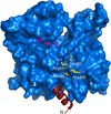
Structure of the W domain of the NPF protein WAVE2 (red) bound to actin (blue). Actin subdomains 1–4 are labeled. The W domain consists of an N-terminal amphiphilic helix that binds in the so-called hydrophobic or target-binding cleft (Dominguez, 2004) between actin subdomains 1 and 3, followed by a C-terminal extended region, featuring the conserved LKKT(V) motif (449LRRV452 in WAVE2). This and other W-actin structures were determined as ternary complexes with DNase I, which prevents polymerization (Chereau et al., 2005). For simplicity, DNase I is not shown. A color version of this figure is available online.
Figure 3
Figure 3
Actin filament nucleation by the Arp2/3 complex and NPFs. (A) The Arp2/3 complex consists of seven proteins, including the actin related proteins Arp2 and Arp3, and subunits ARPC1 to 5 (labeled 1 to 5). By itself, the complex has low nucleation activity, but it is activated by Nucleation Promoting Factors (NPFs) (Goley and Welch, 2006; Pollard, 2007). NPFs are large multi-domain proteins. The figure illustrates a prototypical NPF protein, characterized by the presence of regulatory/localization domains, a Pro-rich region, and a WCA region that can have between one and three W domains. Two of the W domains are colored red/gray (striped) to indicate that their presence is not absolutely necessary for Arp2/3 complex activation. WCA is the smallest fragment capable of catalyzing the formation of a polymerization nucleus, consisting of the two Arps and one to three actin subunits, as well as a conformational change within Arp2/3 complex that allows for monomer addition to the branch and binding of the nucleus to the side of a preexisting actin filament (mother filament). The new filament (or branch) grows from the barbed ends of the Arps at a 70° angle with respect to the mother filament. (B) Structure of inactive Arp2/3 complex (Nolen and Pollard, 2007; Robinson et al., 2001). Subdomains 1 and 2 of Arp2 are disordered in the structures, but were added here by analogy with actin. The Arp2/3 complex subunits are colored according to the diagram of part A. (C) SAXS-derived model of WCA-actin-Arp2/3 complex (Boczkowska et al., 2008). The orientation is the same as in part B. The mother and branch filaments are shown for reference, although this work did not address branch assembly. Note that Arp2 moved up compared to its location in part B. This study placed the first actin subunit of the branch (bound to the W domain) at the barbed end of Arp2. The position of the crosslinked W domain, which is known precisely from its crystal structure with actin (Chereau et al., 2005) (see Figure 2), imposes constraints on the location of the C motif. Thus, the hydrophobic cleft of Arp2 is in the path of the CA polypeptide as it progresses toward the Arp2/3 complex. The helical portion of the C motif may thus bind in this cleft, as supported by sequence similarity with W (see Figure 4C). The position of the A motif (pink) is less well constrained, but this model would be consisting with it binding at the interface between Arp3 and ARPC3, as suggested by biochemical studies (Kelly et al., 2006; Kreishman-Deitrick et al., 2005; Pan et al., 2004; Weaver et al., 2002; Zalevsky et al., 2001). A color version of this figure is available online.
Figure 4
Figure 4
Relationship between tandem W-based filament nucleators, NPFs and Ena/VASP elongation factors. (A) Domain diagram. W and C are both colored red to highlight their relationship. Pro-rich regions (magenta) abound among these proteins, and bind regulatory proteins and profiling-actin complexes, which is the main source of polymerization competent actin in cells. Coiled coils and other oligomerization domains (both known and predicted from sequence analysis) are also common (green). In Ena/VASP, the WASP-Homology domain 1 (WH1), W and C regions are respectively known as EVH1 (Ena/VASP homology 1), GAB and FAB (G- and F-actin binding) domains, but despite their different names these domains are related to their N-WASP counterparts. Ena/VASP also has an Acidic region after the C motif, albeit less acidic than in NPFs and lacking the key tryptophan residue. An ubiquitin-like segment in Cobl (a potential Ras-binding site), a snare-like helix in WAVE2 (a potential multi-protein association site), a spectrin-like antiparallel dimerization motif in JMY and a globular dimerization domain in VopL/VopF are all predicted by bioinformatics analysis, but had not been previously reported. The third W domain of Spire (red/magenta) is non-canonical; it contains various Pro residues and, interestingly, occupies the position of the Pro-rich linker-2 of Cobl and VopL/VopF. (B) Proposed nucleation mechanisms of Spire (Quinlan et al., 2005) and Cobl (Ahuja et al., 2007). The lengths of the linkers determine whether neighboring W domains bridge actin subunits along a single strand (Spire) or across strands (Cobl) of the actin filament (i.e. long- vs short-pitch nuclei), which may in turn determine the nucleation activities of these proteins. Intriguingly, VopL has three W domains and strong nucleation activity like Cobl, but its shorter linker-2 would be inconsistent with stabilization of a short-pitch nucleus. (C) Sequence alignment of the W and CA regions of the proteins shown in part A. Conserved residues are colored according to their chemical properties. Uniprot codes: Spire (Drosophila melanogaster, Q9U1K1); Cobl (mouse, Q5NBX1); VopL (Vibrio parahaemolyticus, Q87GE5); JMY (mouse, Q9QXM1); N-WASP (mouse, Q91YD9); WAVE2 (human, Q9Y6W5); VASP (human, P50552); Evl (human, Q9UI08). A color version of this figure is available online.
Figure 5
Figure 5
Model of nucleation by NPFs-Arp2/3 complex (see text for details). A color version of this figure is available online.
Figure 6
Figure 6
Domain organization of Lmod and proposed nucleation mechanism. (A) Domain organization of Lmod compared to Tmod. The first ~340aa of Lmod are ~40% identical to Tmod, a pointed end capping protein in muscles. The N-terminal portion of Tmod is unstructured, except for three helical segments involved in binding tropomyosin (TM) and actin. Tmod has a second actin-binding site within the C-terminal Leu-rich repeat (LRR) domain (Fowler et al., 2003; Krieger et al., 2002). Lmod shares this domain organization, but has only one of the two TM-binding sites. More importantly, Lmod has a ~150aa C-terminal extension featuring a third actin-binding site, a W domain, a basic patch and other predicted motifs. (B) Proposed nucleation mechanism; Lmod stabilizes a trimeric actin seed that grows from the barbed end. Importantly, Lmod’s nucleation activity and localization are both modulated by interaction with TM (Chereau et al., 2008). A color version of this figure is available online.
Figure 7
Figure 7
Tβ4 and profilin help maintain the pool of monomeric actin in cells. (A) Model of the structure of the complex of Tβ4-actin. This model was generated by combining the structures of W-actin (Chereau et al., 2005) with that of a complex of actin with the C-terminal half of Tβ4 crystallized as a hybrid with gelsolin segment 1 (Irobi et al., 2004). (B) Structure of the ternary complex of profilin-actin with human VASP fragment Gly-202 to Ser-244 (Ferron et al., 2007). This fragment of VASP includes the last Pro-rich profilin-binding site and the G-actin-binding (GAB) domain, which is related to the W domain (see Figure 2 and Figure 4). The Gly-rich linker between these two domains (213QGPGGGGAG221) was not visualized in the electron density map and is shown as a discontinuous line. A color version of this figure is available online.

Similar articles

Cited by

References

    1. Aguda AH, Burtnick LD, Robinson RC. The state of the filament. EMBO Rep. 2005;6:220–226. - PMC - PubMed
    1. Ahuja R, Pinyol R, Reichenbach N, Custer L, Klingensmith J, Kessels MM, Qualmann B. Cordon-Bleu Is an Actin Nucleation Factor and Controls Neuronal Morphology. Cell. 2007;131:337–350. - PMC - PubMed
    1. Ammer AG, Weed SA. Cortactin branches out: roles in regulating protrusive actin dynamics. Cell Motil Cytoskeleton. 2008;65:687–707. - PMC - PubMed
    1. Auerbuch V, Loureiro JJ, Gertler FB, Theriot JA, Portnoy DA. Ena/VASP proteins contribute to Listeria monocytogenes pathogenesis by controlling temporal and spatial persistence of bacterial actin-based motility. Mol Microbiol. 2003;49:1361–1375. - PubMed
    1. Bachmann C, Fischer L, Walter U, Reinhard M. The EVH2 domain of the vasodilator-stimulated phosphoprotein mediates tetramerization, F-actin binding, and actin bundle formation. J Biol Chem. 1999;274:23549–23557. - PubMed

Publication types

Substances

LinkOut - more resources