. 2010 May;25(5):1103-16.
doi: 10.1359/jbmr.091036.

Transfer, analysis, and reversion of the fibrous dysplasia cellular phenotype in human skeletal progenitors

Affiliations

Transfer, analysis, and reversion of the fibrous dysplasia cellular phenotype in human skeletal progenitors

Stefania Piersanti et al. J Bone Miner Res. 2010 May.

Abstract

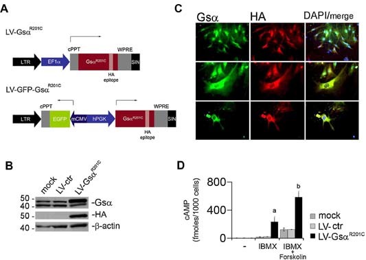
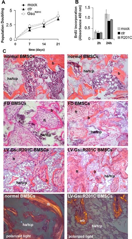
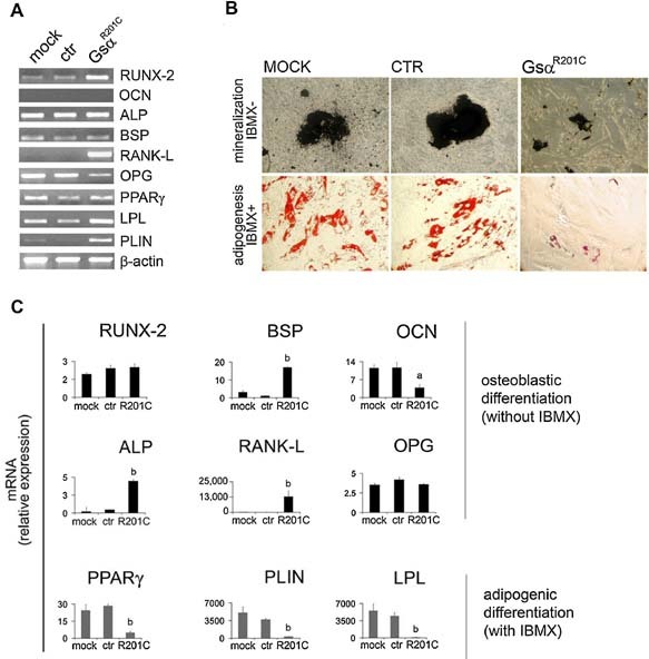
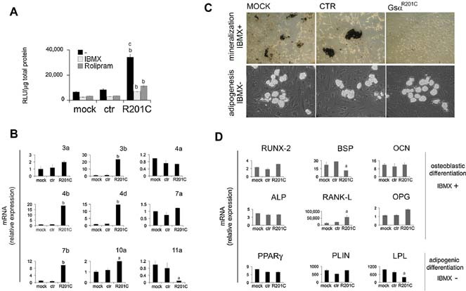
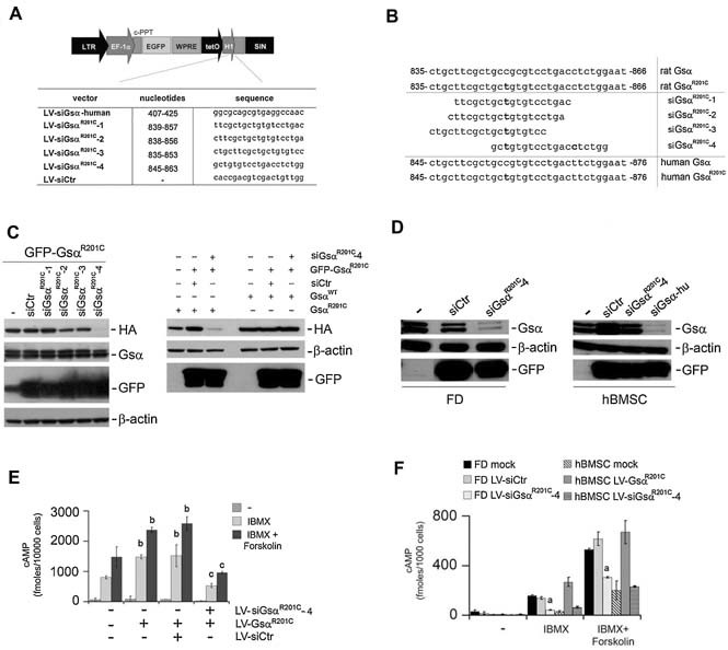
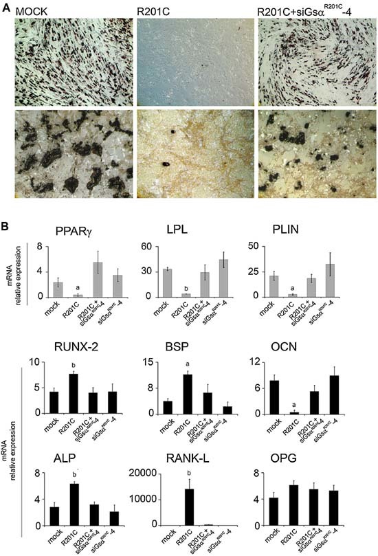
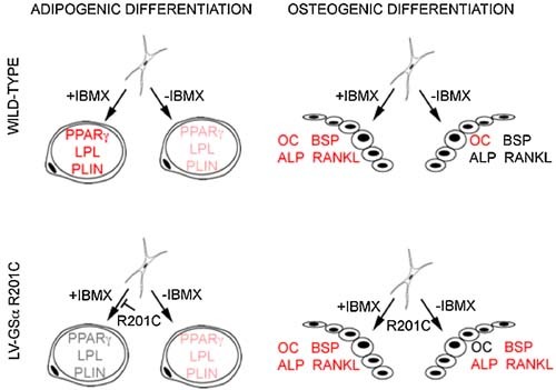
Human skeletal progenitors were engineered to stably express R201C mutated, constitutively active Gs alpha using lentiviral vectors. Long-term transduced skeletal progenitors were characterized by an enhanced production of cAMP, indicating the transfer of the fundamental cellular phenotype caused by activating mutations of Gs alpha. Like skeletal progenitors isolated from natural fibrous dysplasia (FD) lesions, transduced cells could generate bone but not adipocytes or the hematopoietic microenvironment on in vivo transplantation. In vitro osteogenic differentiation was noted for the lack of mineral deposition, a blunted upregulation of osteocalcin, and enhanced upregulation of other osteogenic markers such as alkaline phosphatase (ALP) and bone sialoprotein (BSP) compared with controls. A very potent upregulation of RANKL expression was observed, which correlates with the pronounced osteoclastogenesis observed in FD lesions in vivo. Stable transduction resulted in a marked upregulation of selected phosphodiesterase (PDE) isoform mRNAs and a prominent increase in total PDE activity. This predicts an adaptive response in skeletal progenitors transduced with constitutively active, mutated Gs alpha. Indeed, like measurable cAMP levels, the differentiative responses of transduced skeletal progenitors were profoundly affected by inhibition of PDEs or lack thereof. Finally, using lentiviral vectors encoding short hairpin (sh) RNA interfering sequences, we demonstrated that selective silencing of the mutated allele is both feasible and effective in reverting the aberrant cAMP production brought about by the constitutively active Gs alpha and some of its effects on in vitro differentiation of skeletal progenitors.


Figures

Figure 1
Figure 1
Lentiviral transfer of GsαR201C mutation in hBMSCs. (A) LV vectors used for GsαR201C transfer. LV‐EF‐GsαR201C encodes the GsαR201C protein (HA‐tagged), controlled by the EF‐1α promoter. The bidirectional LV vector (LV‐BD‐GsαR201C) encodes GsαR201C and eGFP under the control of the PGK and CMV minimal promoters, respectively. (B) Western blot analysis of GsαR201C expression in LV‐EF‐GsαR201C‐transduced hBMSCs. Immunoblotting for HA demonstrates the specific signal for the mutated Gsα protein. Immunoblotting for Gsα demonstrates that mock‐treated and empty vector (LV‐Ctr)–transduced cells express comparable amounts of the 48‐ and 43‐kDa isoforms of Gsα (Gsα‐long and Gsα‐short, respectively) resulting from alternative splicing of exon 3. A specific increase of Gsα‐long is observed in cells transduced with LV‐EF‐GsαR201C. (C) GsαR201C expression in hBMSCs is detected by immunofluorescence, as indicated by HA immunolabeling. (D) Intracellular cAMP levels were measured in control or LV‐EF‐GsαR201C‐transduced hBMSCs. In the presence of IBMX (1 mM) alone or of IBMX and forskolin (10 µM), significantly higher levels of cAMP are observed in GsαR201C‐expressing cells compared with control cells. Data from five separate experiments in duplicate are expressed as mean ± SD. a p < .05 versus mock and LV‐ctr; b p < .01 versus mock and ctr.
Figure 2
Figure 2
Effect of GsαR201C on hBMSC proliferation and in vivo differentiation. Analysis of cell proliferation in transduced hBMSCs expressed as (A) population doublings or (B) BrdU incorporation for 2 and 24 hours. The presence of mutated GsαR201C does not alter BMSC proliferation. Data from three experiments are expressed as means ± SD. (C) In vivo transplantation of normal, FD‐derived (R201C) and LV‐ GsαR201C‐transduced hBMSCs. Note the formation of abundant bone (b) on hydroxyapatite (ha/tcp, hydroxyapatite/tricalcium phosphate), hematopoietic tissue (hem, meg = megakaryocytes), and adipocytes (ad) in control transplants (upper panels). Transplants of FD‐derived BMSCs and transduced BMSCs are recognized by the reduced amount of bone and the fibrous tissue (ft) devoid of hematopoietic cells and adipocytes filling the marrow space. Polarized light microscopy demonstrates that normal BMSCs formed lamellar bone (lb), whereas transduced BMSCs formed woven bone (wb).
Figure 3
Figure 3
GsαR201C effect on the adipogenic and osteogenic markers in undifferentiated and in vitro differentiated hBMSCs. (A) Adipogenic and osteogenic marker quantification in GsαR201C‐transduced hBMSCs. RT‐PCR analysis was carried out in undifferentiated uninfected and LV‐ctr‐ or LV‐GsαR201C‐infected hBMSCs. Significant activation of RANKL is detected in LV‐GsαR201C‐infected hBMSCs. (B) Adipogenic and osteogenic differentiation of GsαR201C‐transduced hBMSCs. hBMSCs were cultured in osteogenic and adipogenic medium, and mineralization nodules or adipocyte‐like cells were revealed with Von Kossa and oil red O staining, respectively. Both in vitro osteogenic differentiation and in vitro adipogenic differentiation of hBMSC are altered in GsαR201C‐transduced cells. (C) qPCR of osteogenic and adipogenic markers analyzed in differentiated hBMSCs. Abnormal differentiation was observed in GsαR201C‐transduced cells compared with mock‐transduced and control cells. Data from four experiments in duplicates are expressed as means ± SD. a p < .05 versus mock and ctr; b p < .01 versus mock and ctr.
Figure 4
Figure 4
Activity, mRNA expression, and phenotypic effects of PDEs in mutation‐transduced hBMSCs. (A) PDE activity was assayed in hBMSCs, mock‐treated or transduced with LV‐Ctr or LV‐ EF‐GsαR201C, using 10 µM of cAMP as a substrate, in the presence or absence of either IBMX (1 mM) or Rolipram (20 µM). b p < .01 versus equally treated mock and ctr cells; c p < .01 versus mutation‐transduced, IBMX‐ and rolipram‐treated cells. (B) qPCR analysis of the expression of PDE isoforms involved in cAMP degradation. Significant increase of expression was detected for PDE‐3b, ‐4b, ‐4d, and ‐7b isoforms in mutation‐transduced cells compared with mock‐ and LV‐Ctr‐treated cells. Values represent the mean ± SD of three experiments in duplicate. a p < .05 versus mock and ctr; b p < .01 versus mock and ctr. (C) hBMSCs were cultured in osteogenic medium supplemented with IBMX (0.5 mM) or adipogenic medium without IBMX. Mineralization nodules or adipocyte‐like cells were revealed with Von Kossa and oil red O staining, respectively. (D) PDE inhibition in osteogenic medium caused a significant upregulation of osteogenic markers in mock‐transduced and control cells at the same levels of GsαR201C. Adipogenesis in the absence of IMBX leads to a modest increase of adipogenic markers to the same extent in control and GsαR201C cells. a p < .05 versus mock and ctr.
Figure 5
Figure 5
Specific and stable LV‐mediated silencing of GsαR201C in normal and FD hBMSCs. (A) Schematic representation of the LV vectors expressing the Gsα RNA‐interfering sequences under the control of the H1 promoter and eGFP as a marker. (B) Design of different RNA‐interfering sequences directed to human exon 1 and human/rat GsαR201C exon 8. (C) Left panel: HeLa cells were first infected with the bidirectional LV‐GFP‐GsαR201C and subsequently superinfected with the different LV‐siGsα vectors as indicated. Specific knockdown of mutated Gsα was obtained only with LV‐siGsαR201C‐4. Right panel: HeLa GFP‐GsαR201C/siCtr or HeLa GFP‐GsαR201C/siGsαR201C‐4 were superinfected with LV‐EF1α‐GsαR201C or LV‐EF1α‐GsαWT. A specific downregulation of the added‐in mutated R201C Gsα was observed. In contrast, robust expression of the added‐in GsαWT was observed. (D) Left panel: Highly mutated in R201C, FD‐derived hBMSCs were mock infected or treated with LV‐siCtr or LV‐siGsαR201C‐4 and after 20 days of culture, the cells were collected for Western blot analysis. A decrease in both the long and short endogenous form of Gsα was observed with LV‐siGsαR201C‐4. Right panel: Normal hBMSCs were untreated or treated with LV‐siCtr, LV‐siGsαR201C‐4, and LV‐siGsα human exon 1 (hu). The 20 days after infection, cells were processed for Western blotting. Both Gsα endogenous WT isoforms (long and short Gsα) were efficiently suppressed with siGsα exon 1 but remained efficiently expressed with specific siGsαR201C‐4. (E) cAMP assay on HeLa LV‐GFP‐GsαR201C/siGsαR201C‐4 demonstrates the capacity to restore the cAMP levels similar to that of the basal levels. Data are means ± SD of n = 3 experiments in duplicate. b p < .01 versus untransduced HeLa equally treated; c p < .01 versus LV‐GsαR201C and LV‐GsαR201C/LV‐siCtr cells equally treated. (F) cAMP assay on transduced FD‐derived BMSCs and normal hBMSCs. A significant reduction of cAMP levels, comparable with that recorded in normal hBMSC, was observed in FD‐LV‐siGsαR201C‐4. a p < .05 of FD‐LV‐siGsαR201C‐4 versus FD mock, FD‐LV‐siCtr, and hBMSC‐GsαR201C.
Figure 6
Figure 6
Specific lentivirus‐directed RNA interference of constitutively active Gsα restores in vitro differentiation of LV‐GsαR201C‐transduced skeletal progenitors. (A) Restoration of in vitro adipogenesis (top, oil red O) and mineralization (bottom, von Kossa staining) in BMSCs that were first transduced with LV‐GsαR201C and then superinfected with LV‐siGsαR201C‐4. (B) Restoration of expression of mRNAs for adipogenic and osteogenic marker, qPCR analysis. Knockdown of mutated Gsα by LV‐siGsαR201C‐4 in previously GsαR201C‐transduced cells reestablishes the markers expression at the same levels as seen in mock cells. siGsαR201C‐4 does not affect the differentiation of untransduced cells. Data from four experiments in duplicate are expressed as means ± SD. a p < .05 versus mock, LV‐GsαR201C plus LV‐siGsαR201C‐4, and LV‐siGsαR201C‐4; b p < 0.01 versus mock, LV‐GsαR201C plus LV‐siGsαR201C‐4 and LV‐siGsαR201C‐4. No significant difference in expression levels occurs among mock and LV‐GsαR201C plus LV‐siGsαR201C‐4, mock, and LV‐siGsαR201C‐4 or LV‐GsαR201C plus LV‐siGsαR201C‐4 and LV‐siGsαR201C‐4.
Figure 7
Figure 7
Schematic representation of the interplay of Gsα and PDE activity in the regulation of in vitro adipogenic and osteogenic differentiation of BMSCs. Inhibition of PDE (IBMX) results in marked enhancement of upregulation of adipogenic markers in BMSCs induced to in vitro adipogenesis. Constitutive activity of Gsα (R201C) results in a less prominent but clear‐cut enhanced expression of adipogenic markers. Inhibition of PDE in the presence of constitutively active Gsα, in contrast, results in the block of adipogenic differentiation. In BMSCs induced to in vitro osteogenic differentiation, addition of IBMX enhances the upregulation of BSP, ALP, and RANKL. Enhanced expression of the same markers is also observed in the absence of IBMX as an effect of constitutive activity of Gsα. Magnitude of enhancement is represented schematically by red, light red, and gray character colors in the notation of the different markers.

References

    1. Weinstein LS, Shenker A, Gejman PV, Merino MJ, Friedman E, Spiegel AM. Activating mutations of the stimulatory G protein in the McCune‐Albright syndrome. N Engl J Med 1991; 325: 1688–1695. - PubMed
    1. Shenker A, Weinstein LS, Moran A, et al. Severe endocrine and nonendocrine manifestations of the McCune‐Albright syndrome associated with activating mutations of stimulatory G protein GS. J Pediatr 1993; 123: 509–518. - PubMed
    1. Masters SB, Miller RT, Chi MH, et al. Mutations in the GTP‐binding site of GSα alter stimulation of adenylyl cyclase. J Biol Chem 1989; 264: 15467–15474. - PubMed
    1. Levis MJ, Bourne HR. Activation of the α subunit of Gs in intact cells alters its abundance, rate of degradation, and membrane avidity. J Cell Biol 1992; 119: 1297–1307. - PMC - PubMed
    1. Bourne HR, Landis CA, Masters SB. Hydrolysis of GTP by the α‐chain of Gs and other GTP‐binding proteins. Proteins 1989; 6: 222–230. - PubMed

Publication types

MeSH terms

Substances