. 2010 Jan;8(1):185-93.
doi: 10.1111/j.1538-7836.2009.03662.x. Epub 2009 Oct 23.

Cleaved high molecular weight kininogen inhibits tube formation of endothelial progenitor cells via suppression of matrix metalloproteinase 2

Affiliations

Cleaved high molecular weight kininogen inhibits tube formation of endothelial progenitor cells via suppression of matrix metalloproteinase 2

Y Wu et al. J Thromb Haemost. 2010 Jan.

Abstract

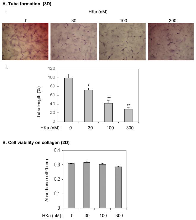
Background and objective: Endothelial progenitor cells (EPCs) contribute to postnatal neovascularization, thus promoting wide interest in their therapeutic potential in vascular injury and prevention of their dysfunction in cardiovascular diseases. Cleaved high molecular weight kininogen (HKa), an activation product of the plasma kallikrein-kinin system (KKS), inhibits the functions of differentiated endothelial cells including in vitro and in vivo angiogenesis. In this study, our results provided the first evidence that HKa is able to target EPCs and inhibits their tube forming capacity.

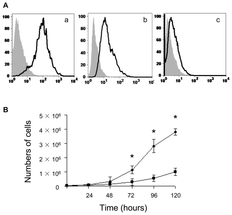
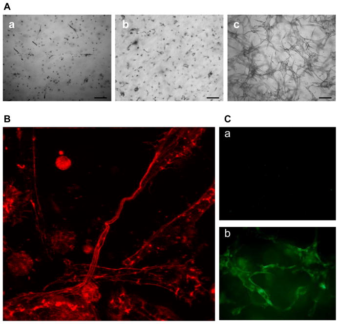
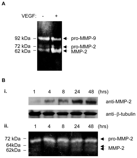
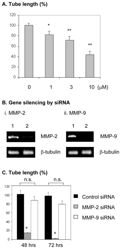
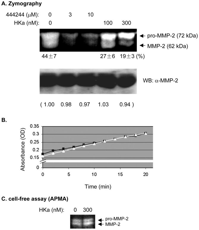
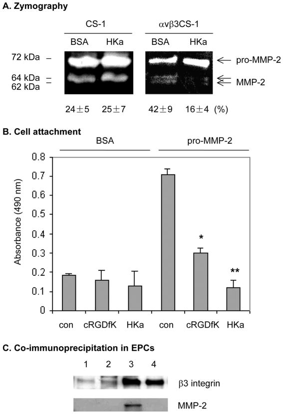
Methods and results: We determined the effect of HKa on EPCs using a three-dimensional vasculogenesis assay. Upon stimulation with vascular endothelial growth factor (VEGF) alone, EPCs formed vacuoles and tubes, and differentiated into capillary-like networks. As detected by gelatinolytic activity assay, VEGF stimulated secretion and activation of matrix metallopeptidase 2 (MMP-2), but not MMP-9, in the conditioned medium of 3D culture of EPCs. Specific inhibition or gene ablation of MMP-2, but not MMP-9, blocked the vacuole and tube formation by EPCs. Thus, MMP-2 is selectively required for EPC vasculogenesis. In a concentration-dependent manner, HKa significantly inhibited tube formation by EPCs and the conversion of pro-MMP-2 to MMP-2. Moreover, HKa completely blocked the association between pro-MMP-2 and alphavbeta3 integrin, and its inhibition of MMP-2 activation was dependent on the presence of alphavbeta3 integrin. In a purified system, HKa did not directly inhibit MMP-2 activity.

Conclusions: HKa inhibits tube forming capacity of EPCs by suppression of MMP-2 activation, which may constitute a novel link between activation of the KKS and EPC dysfunction.


Figures

Figure 1
Figure 1. Immunophenotyping and high proliferation capacity of EPCs
(A) Immunophenotyping of cell-surface by flow cytometry (a, CD-31; b, VE-cadherin; c, AC133). Shown are representative data from 4 independent experiments. Isotype controls are indicated in gray shadow. (B) Growth of EPCs (diamonds) and HAECs (squares) in EGM-2 was evaluated in equivalently seeded in vitro. *p<0.01.
Figure 2
Figure 2. EPCs exhibit potent capacity to form tubular structures
(A) HAECs (a), HUVECs (b) and EPCs (c) were cultured in the presence of VEGF (25 ng/mL) in 3D collagen gel for 48 hours. Bar = 100 μm. The images are representative of three independent experiments. (B) Formation of capillary-like networks by EPCs. After culture for 48 hours, EPCs in collagen gel were stained with rhodamine-phalloidin and visualized by confocal microscope (400×). The image represents a maximum projection of the stacked optical sections. (C) Tube structure of EPCs in 3D was visualized by immunostaining with anti-vWF (b) or control IgG (a) plus Alexa Fluor@ 488-GαM antibody.
Figure 3
Figure 3. MMP-2, but not MMP-9, is activated during VEGF-stimulated EPC differentiation
(A) Zymography assay. After EPCs embedded in collagen gel were cultured with or without 25 ng/mL of VEGF for 24 hours, the gelatinolytic activity in the conditioned culture media was analyzed by zymography. (B) VEGF (25 ng/ml)-stimulated expression and secretion of MMP-2 is time-dependent. The expression of cytosolic MMP-2 in EPCs as determined by Western-blot with β-tubulin serving as a loading control (i); Zymography of the conditioned culture media (ii). The results are representative of three independent experiments.
Figure 4
Figure 4. MMP-2 activity and expression is selectively required for EPC differentiation in response to VEGF stimulation
(A) EPCs were cultured in 3D collagen gel for 48 hours in the presence o f 25 ng/mL VEGF plus specific MMP-2 inhibitor (444244) at the indicated concentrations. As described in the Methods, the sum of the tube length in six digital images per well was indicated as micrometer per square millimeter. Results are given as percentage of tube length/area compared with the control (0 μM) (which was set as 100%). Experiments were done in triplicates. * p< 0.05; ** p<0.01. (B) Gene silencing of MMP-2 (i) and MMP-9 (ii). The knockdown efficiency by siRNA was evaluated by RT-PCR. Lane 1, control non-silencing siRNA, lane 2, MMP-2 or MMP-9 siRNA. (C) After transfection with siRNA, EPCs in 3D collagen gel were cultured with VEGF for 48 and 72 hours, respectively. Experiments at each time point were done in triplicates. Quantitation of tube length was done as described in A. Results are given as percentage of tube length/area compared with the control group at 48 hours (which was set as 100%). *p<0.0001; n.s: not significant, compared with control.
Figure 5
Figure 5. HKa inhibits EPC differentiation without affecting cell viability
(A) i. Morphogenesis of EPCs in collagen gel. EPCs were suspended in EBM-2 containing 25 μM ZnCl2 and incubated with or without HKa on ice for 20 minutes. After mixture with the collagen gel solution, EPCs were cultured in the presence of VEGF (25 ng/mL) for 48 hours. ii. Tube length was measured and analyzed as described in the Figure 4A legend. Experiments were done in triplicate. * p<0.01; **p<0.005. (B) EPCs were cultured on collagen-coated plate in the presence of VEGF (25 ng/ml) plus HKa at the indicated concentrations for 48 hours (n=4). The cell number was determined as described in the Methods.
Figure 6
Figure 6. HKa inhibits the conversion of proMMP-2 to MMP-2
(A) EPCs were cultured in 3D collagen matrices in the presence of VEGF (25 ng/mL) plus MMP-2 Inhibitor I (444244) or HKa as indicated. The conditioned medium was collected for zymography assay (upper panel) and anti-MMP-2 Western-blot (lower panel). Intensity of pro-MMP-2 and MMP-2 bands in zymography gel was quantified using software Quantity One, version 4.1.1 (Bio-Rad) after background subtraction. The number under the zymography gel indicates the band intensity ratio of MMP-2/(MMP-2 + pro-MMP-2) (%), average ± SEM, n=3. (B) Gelatinase activity assay. Recombinant human MMP-2 catalytic domain was incubated with a thiopeptide as a chromogenic substrate in the presence of 0.1% BSA or 300 nM HKa. Absorbance values were read as described in the Methods. The results are representative of three independent experiments. (■, BSA; △, HKa). (C) Recombinant pro-MMP-2 was incubated with APMA with 0 nM or 300 nM HKa at 37°C for 60 min. Native and processed MMP-2 were detected by zymography.
Figure 7
Figure 7. HKa inhibition of MMP-2 activation is dependent on αvβ3 integrin
(A) CS-1 and αvβ3CS-1 cells were transfected with full length human MMP-2 cDNA for 48 hours. After incubation with 0.1% BSA or 300 nM HKa in serum-free medium overnight, the conditioned medium was collected for zymography assay. The band intensity was calculated as described in the Figure 4B legend and the number shown under the gel represents average ± SEM, n=3. (B) Cell attachment. The 96-well cell culture plates were coated with 0.3% BSA and 10 μg/mL recombinant proMMP-2 protein. The αvβ3CS-1 cells (5×10⁴ cells/well) were cultured at 37°C in the presence of 0.1% BSA, 10 μM cRGDfK, or 300 nM HKa for 2 hours. After unattached cells were removed, the attached cell number was determined as described in the Methods. Experiments were done in triplicate. *p<0.05; **p<0.01. (C) Effect of HKa on αvβ3 integrin- MMP-2 complex in EPCs. As described in the legend to Figure 5A, EPCs were incubated in a 3- D gel with (lane 2 and lane 4) or without (lane 1 and lane 3) 300 nM HKa for 48 hours. Cells were harvested by the addition of extraction buffer. Cell lysates were subjected to immunoprecipitation with mouse IgG (lane 1 and lane 2) or LM609 (lane 3 and 4). The immunoprecipitates were probed by antibodies against β3 integrin and MMP-2, respectively. Data are representative of 3 independent experiments.

References

    1. Yoder MC, Mead LE, Prater D, Krier TR, Mroueh KN, Li F, Krasich R, Temm CJ, Prchal JT, Ingram DA. Redefining endothelial progenitor cells via clonal analysis and hematopoietic stem/progenitor cell principals. Blood. 2007;109:1801–9. - PMC - PubMed
    1. Prater DN, Case J, Ingram DA, Yoder MC. Working hypothesis to redefine endothelial progenitor cells. Leukemia. 2007;21:1141–9. - PubMed
    1. Werner N, Nickenig G. Endothelial progenitor cells in health and atherosclerotic disease. Annals of Medicine. 2007;39:82–90. - PubMed
    1. Melero-Martin JM, Khan ZA, Picard A, Wu X, Paruchuri S, Bischoff J. In vivo vasculogenic potential of human blood-derived endothelial progenitor cells. Blood. 2007;109:4761–8. - PubMed
    1. Colman RW, Schmaier AH. Contact System: A Vascular Biology Modulator With Anticoagulant, Profibrinolytic, Antiadhesive, and Proinflammatory Attributes. Blood. 1997;90:3819–43. - PubMed

Publication types

MeSH terms