. 2009 Oct 29;64(2):165-72.
doi: 10.1016/j.neuron.2009.09.016.

Combined intrinsic and extrinsic neuronal mechanisms facilitate bridging axonal regeneration one year after spinal cord injury

Affiliations

Combined intrinsic and extrinsic neuronal mechanisms facilitate bridging axonal regeneration one year after spinal cord injury

Ken Kadoya et al. Neuron. .

Abstract

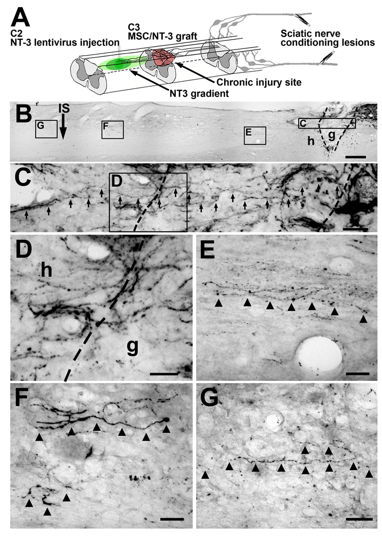
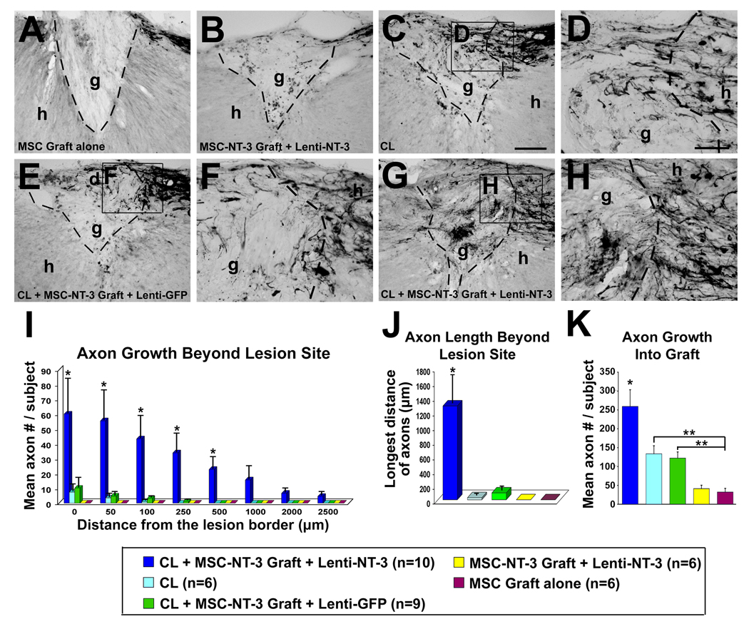
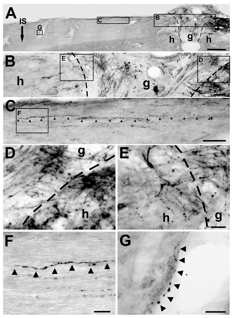
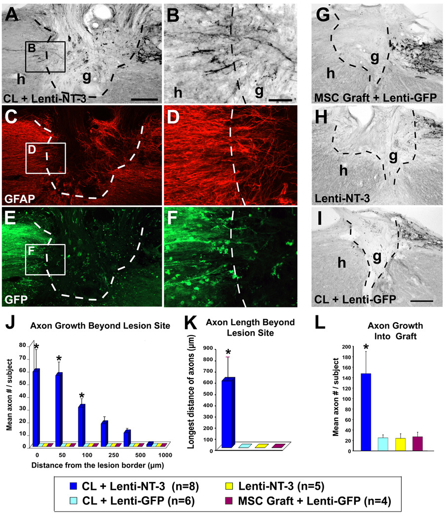
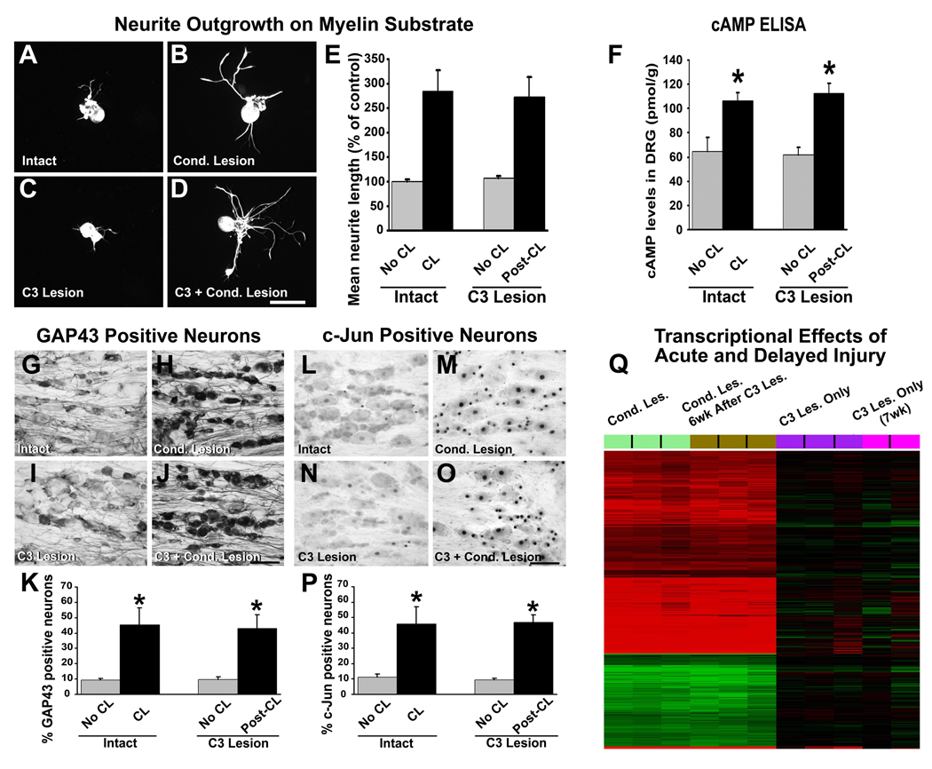
Despite advances in promoting axonal regeneration after acute spinal cord injury (SCI), elicitation of bridging axon regeneration after chronic SCI remains a formidable challenge. We report that combinatorial therapies administered 6 weeks, and as long as 15 months, after SCI promote axonal regeneration into and beyond a midcervical lesion site. Provision of peripheral nerve conditioning lesions, grafts of marrow stromal cells, and establishment of NT-3 gradients supports bridging regeneration. Controls receiving partial components of the full combination fail to exhibit bridging. Notably, intraneuronal molecular mechanisms recruited by delayed therapies mirror those of acute injury, including activation of transcriptional activators and regeneration-associated genes. Collectively, these findings provide evidence that regeneration is achievable at unprecedented postinjury time points.


Figures

Figure 1
Figure 1
Sensory axons regenerate beyond the lesion site when treatments influencing both neuron-intrinsic and environmental mechanisms are applied 6 weeks after the original SCI. (A) Injury model and delayed combination therapy. Dorsal column sensory axons were completely transected at C3. Six weeks or 15 months later, L4-6 DRG neurons were conditioned by sciatic nerve crush. One week later, MSCs were injected into the lesion site, and NT-3-expressing lentivirus was injected 2.5mm rostral to the lesion site. Animals survived an additional 6 weeks. (B) Low magnification sagittal overview from subject that received delayed combination therapy. CTB-labeled dorsal column sensory axons are evident approaching lesion/graft site on upper right side of panel. Rostral left, caudal right. g, graft; h, host; IS, lenti-NT-3 injection site 2.5mm rostral to lesion border. Scale bar 200µm. Areas of higher magnification are indicated in boxed regions. (C) Higher magnification of graft: axons indicated by arrows course through graft in irregular trajectories; several axons cross rostral interface with host (dashed line in box C) to enter host spinal cord beyond the lesion. (D) High magnification of panel C at interface of graft border with host beyond the lesion, demonstrating sensory axons crossing interface. Notably, axons cross at several dorsoventral levels of host/graft interface, not simply at most dorsal or ventral aspects of grafts where spared axons might be mistaken for regenerating axons. (E) Several varicose axons continue to extend 500µm beyond lesion site in host white matter. (F) 2mm and (G) 2.5 mm beyond the lesion, bridging axons remain visible in host white matter. Lesion completeness was confirmed by sectioning medulla, showing absence of CTB labeling (Suppl. Fig. 1). Scale bar C = 40µm; D–G = 20µm.
Figure 2
Figure 2
Penetration of lesion site and quantification of regeneration. Sagittal sections of C3 lesion/graft site, 3 months after original injury. Rostral left, caudal right. (A) Subjects treated only with MSC grafts demonstrate rare penetration of CTB-labeled sensory axons into graft, and no bridging. g, graft; h, host. Lines indicate host/graft interface. (B) Injection of lenti-NT-3 vector beyond lesion site, and NT-3 protein into MSC graft in lesion site, results in slightly enhanced axon penetration of graft but no axonal bridging beyond graft. (C, D) CLs significantly enhance axon penetration into graft, but axons rarely extend beyond the lesion, and never for distances greater than 100μm. D is higher magnification of panel C, showing axonal entry into graft from caudal host/graft interface. (E, F) Treatment with CL and NT-3 protein within MSC graft, and Lenti-GFP injection beyond, fails to support axonal bridging beyond that observed with CL alone. F is higher magnification of panel E, showing axonal entry into graft from caudal host/graft interface. (G, H) As noted in Figure 1, combinatorial treatment with CL and NT-3 gradients beyond the lesion support axon entry into graft and significant axonal bridging beyond lesion. H is higher magnification of panel G, showing axonal entry into graft. (I) Quantification of regeneration beyond lesion/graft site. Animals that received CLs plus NT-3 gradients exhibit significantly more sensory axons regenerating up to 500μm beyond the lesion (Kruskall Wallis test, χ2 p<0.03; Dunn post-hoc tests with Bonferroni correction p<0.005). (J) Similarly, quantification of longest bridging axon per subject demonstrates significant effects of combinatorial treatments (Kruskall Wallis test, χ2 p<0.01; Dunn post-hoc with Bonferroni correction p<0.005). (K) Quantification of axon number within graft demonstrates greatest growth into grafts in subjects receiving CLs and NT-3 (ANOVA p<0.0001; posthoc Fisher’s *p<0.005 compared to all groups). Subjects that received CLs with or without single injections of NT-3 protein in the graft also showed greater axon penetration than subjects with grafts only (posthoc Fisher’s *p<0.05 in both cases). Scale bar A, B, C, E, G = 100 µm; D, F, H= 50 µm.
Figure 3
Figure 3
15 months delayed combinatorial treatment with CLs, NT-3 gradients, and MSC graft in lesion site promotes sensory axonal bridging in chronic SCI. (A) Low magnification sagittal overview. CTB-labeled sensory axons are evident approaching lesion/graft site on upper right side of panel. Rostral left, caudal right. g, graft; h, host; IS, lenti-NT-3 injection site, 2.5 mm rostral to lesion; dashed lines, graft/lesion border. Scale bar 300µm. (B) Higher magnification of graft/lesion region. CTB-labeled sensory axons enter graft (box D) and extend beyond graft into host spinal cord rostral to lesion (box E). (D, E) Higher magnification of panel B, including caudal (D) and rostral (E) interface of graft with host. Sensory axons cross host/graft interface at several dorsoventral levels. (C) High magnification of panel A: arrowheads indicate a varicose axon extending 1200µm beyond lesion site in host white matter. (F) High magnification of panel C. Arrowheads indicate a regenerating, varicose axon. (G) High magnification of panel A demonstrates a regenerating axon 2200µm beyond lesion site in host white matter, extending in close association with blood vessel wall. All lesions were complete, indicated by sectioning of medulla (Suppl. Fig. 1). Scale bar B, C = 50µm; D, E = 20µm, F = 20µm, G = 25µm.
Figure 4
Figure 4
Only subjects undergoing stimulation of both the chronically injured neuron and the lesioned environment exhibit significant axonal bridging, when treatment is initiated 15 months after SCI. (A–F) Sagittal sections of C3 lesion/graft site, 16.5 months after the original injury, triple labeled for CTB (A, B), GFAP (C, D) and GFP (E, F); rostral left, caudal right. In subjects that received CLs, cell grafts and NT-3 vector injections beyond the lesion site 15 months after the original injury, (A, B) CTB-labeled sensory axons extend into and then (B) emerge from the lesion site. (C, D) GFAP labeling outlines the lesion border; regenerating axons extend into and beyond the GFAP-reactive lesion boundary. (E, F) GFP labeling indicates extension of the lentiviral-NT-3 trophic gradient from the site of vector injection to the immediate host/graft interface: axons bridged beyond the lesion site only when lentiviral-NT-3 vector spread to the lesion/graft boundary (see Suppl. Fig. 1). CTB-labeled sensory axons penetrated grafts, but did not bridge beyond the lesion site, in subjects treated with (G) cell grafts alone, (H) lentiviral-NT-3 gradients + cell grafts but no CL, or (I) CLs + cell grafts, but no NT-3 gradient. (J) Quantification of axon growth beyond lesion/graft site. Only animals that received full treatment exhibited sensory axonal regeneration beyond the lesion site. (Kruskall Wallis test, χ2 p<0.003; Dunn post-hoc with Bonferroni correction p<0.008). (K) Longest bridging axon beyond the graft, per subject. (Kruskall Wallis test, χ2 p<0.003; Dunn post-hoc with Bonferroni correction p<0.008) (L) Quantification of axon number within graft demonstrates significantly greater axonal penetration in subjects receiving full treatment (ANOVA p<0.01; post-hoc Fisher’s *p<0.01 comparing combination treatment to all other groups). Scale bar A, C, E = 200µm; B, D, F = 50µm, G, H, I = 200µm.
Figure 5
Figure 5
Delayed CLs increase neuron-intrinsic growth programs. (A–D) Lumbar DRG neurons were cultured on myelin for 48 hours and labeled with NF200. Neurons from (A) naïve controls extend shorter neurites than (B) CLs 1 week prior to isolation (pre-CL). (C) Dorsal column lesions 6 weeks prior to isolation have no influence on neurite extension, whereas (D) CLs applied 6 weeks following C3 lesions (post-CL) significantly enhance neurite extension. Scale bar AD = 80 µm. (E) Quantification of neurite outgrowth as mean percentage of control (A) indicates that both pre-CL and post-CL significantly enhance neurite outgrowth on myelin compared to intact animals and animals that received only C3 lesions (ANOVA p<0.0001; post-hoc Fisher’s * p<0.0001). (F) Measurement of cAMP levels by ELISA 1 week after either pre- or post-CL demonstrated significant elevations over basal levels (ANOVA p<0.003; post-hoc Fisher’s * p<0.001); C3 lesions alone have no effect. (G–K) Immunolabeling for GAP43 in lumbar DRG neurons shows weak immunoreactivity in (G) DRGs from naïve control animals. (H) Pre-CLs increase intensity and number of labeled neurons. (I) C3 lesions alone placed 6 weeks earlier do not increase GAP43 immunolabeling, whereas (J) post-CLs increase GAP43 labeling to same degree as pre-CL. (K) Quantification of percentage of GAP43-labeled neurons reveals significant increase after pre- and post-CL (ANOVA p<0.01; post-hoc Fisher’s * p<0.01). (L–P) Immunolabeling for c-Jun in lumbar DRG neurons also shows few labeled nuclei in (L) naïve control animals. (M) Pre-CL increases the number and intensity of neurons labeled for c-Jun. (N) C3 lesions alone, 6 weeks earlier, do not increase c-Jun, whereas (O) post-CL 6 weeks after C3 lesions increases c-Jun labeling to same degree as pre-CL. (P) Quantification shows significant increase in percentage of c-Jun-labeled neurons after pre- and post-CLs (ANOVA p<0.003; post-hoc Fisher’s *p<0.005). Scale bar GJ, LO = 80 µm. (Q) Heatmap illustrating hierarchical clustering of gene expression from rat DRG extracted 1 week after pre- or post-CLs, or after C3 lesion alone. Comparisons in each case are made to intact DRGs. Red indicates increased, and green indicates decreased, gene expression. Groups of samples are color-coded. (Light green) Pre-CL upregulates more than 3,000 probes compared to intact DRG. (Brown) Post-CL persistently regulates, with similar intensity, a nearly identical set of probes compared to a pre-CL. In contrast to gene changes observed after CL, a C3 central lesion results in perturbation of remarkably fewer probe sets, whether gene changes are examined acutely after C3 lesion (purple) or seven weeks after C3 injury (pink).

Similar articles

Cited by

References

    1. Azizi SA, Stokes D, Augelli BJ, DiGirolamo C, Prockop DJ. Engraftment and migration of human bone marrow stromal cells implanted in the brains of albino rats--similarities to astrocyte grafts. Proc Natl Acad Sci U S A. 1998;95:3908–3913. - PMC - PubMed
    1. Bunge MB. Bridging areas of injury in the spinal cord. Neuroscientist. 2001;7:325–339. - PubMed
    1. Busch SA, Silver J. The role of extracellular matrix in CNS regeneration. Curr Opin Neurobiol. 2007;17:120–127. - PubMed
    1. Coumans JV, Lin TT, Dai HN, MacArthur L, McAtee M, Nash C, Bregman BS. Axonal regeneration and functional recovery after complete spinal cord transection in rats by delayed treatment with transplants and neurotrophins. J Neurosci. 2001;21:9334–9344. - PMC - PubMed
    1. Fawcett JW. Overcoming inhibition in the damaged spinal cord. J Neurotrauma. 2006;23:371–383. - PubMed

Publication types

MeSH terms