. 2009 Oct 30;16(10):1064-74.
doi: 10.1016/j.chembiol.2009.09.017.

Directed evolution and structural characterization of a simvastatin synthase

Affiliations

Directed evolution and structural characterization of a simvastatin synthase

Xue Gao et al. Chem Biol. .

Abstract

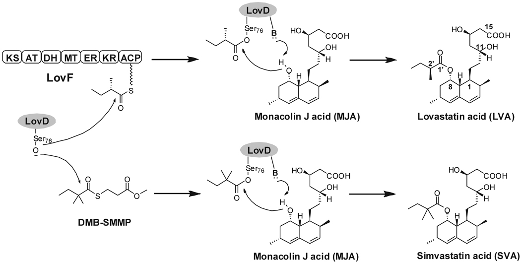
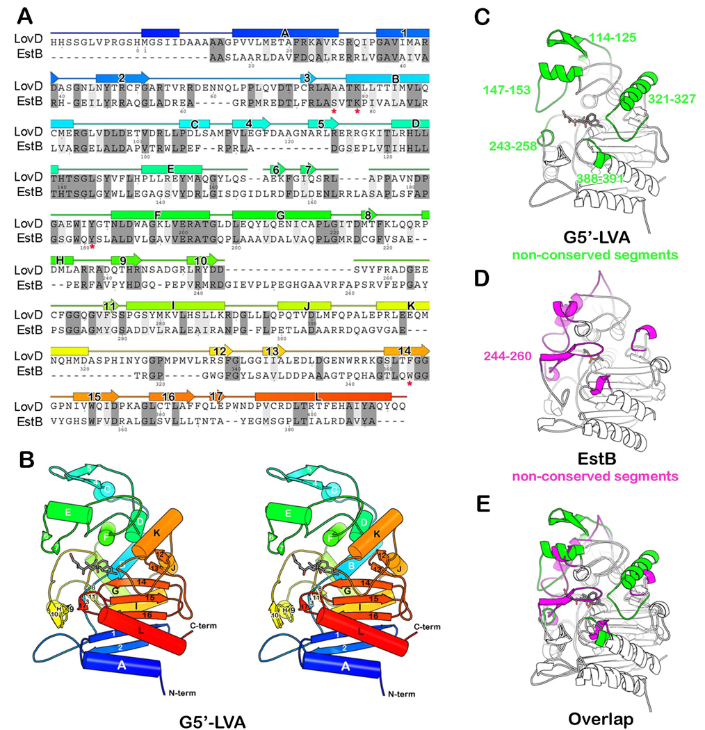
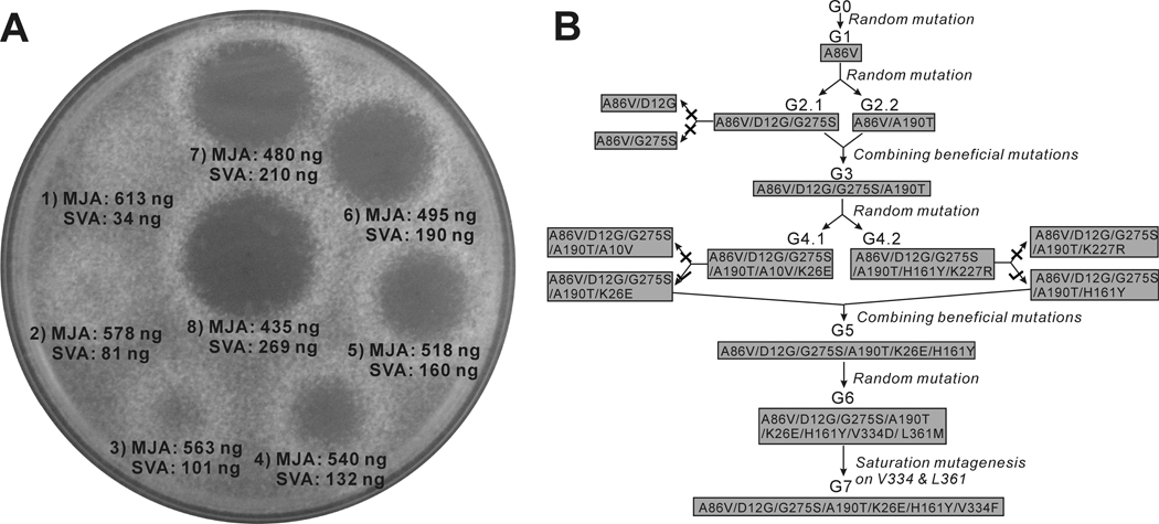
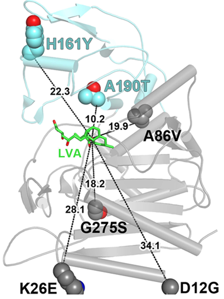
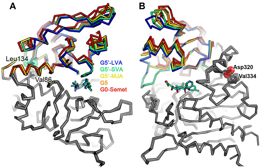
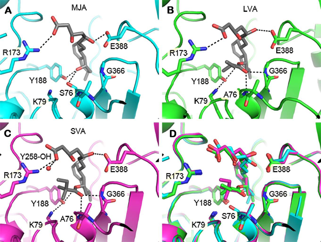
Enzymes from natural product biosynthetic pathways are attractive candidates for creating tailored biocatalysts to produce semisynthetic pharmaceutical compounds. LovD is an acyltransferase that converts the inactive monacolin J acid (MJA) into the cholesterol-lowering lovastatin. LovD can also synthesize the blockbuster drug simvastatin using MJA and a synthetic alpha-dimethylbutyryl thioester, albeit with suboptimal properties as a biocatalyst. Here we used directed evolution to improve the properties of LovD toward semisynthesis of simvastatin. Mutants with improved catalytic efficiency, solubility, and thermal stability were obtained, with the best mutant displaying an approximately 11-fold increase in an Escherichia coli-based biocatalytic platform. To understand the structural basis of LovD enzymology, seven X-ray crystal structures were determined, including the parent LovD, an improved mutant G5, and G5 cocrystallized with ligands. Comparisons between the structures reveal that beneficial mutations stabilize the structure of G5 in a more compact conformation that is favorable for catalysis.


Figures

Figure 1
Figure 1
Reactions catalyzed by LovD. LovD is responsible for converting MJA into LVA via acylation of the α-S-methylbutyrate side chain and can also synthesize SVA using DMB-SMMP as an acyl donor.
Figure 2
Figure 2
The crystal structure of LovD and its relationship to EstB. (A) A structure-based sequence alignment between LovD and EstB. Secondary structure elements assigned from the structure of LovD are shown above the sequence. The colors are ramped from blue at the N-terminus to red at the C-terminus. The active site residues in EstB are indicated by an asterisk “*” below the amino acid. (B) A ribbon diagram showing the G5’-LVA complex. (C) Structure of LovD. Highlighted in green are segments that are not conserved in EstB. These five loops project around the circumference of the active site like the fingers in a catcher’s mitt. (D) Structure of EstB. Highlighted in magenta are segments that are not conserved in LovD. (E) The overlay of LovD and EstB structures. Notably absent from LovD is a loop that covers the active site in EstB (residues 244–260).
Figure 3
Figure 3
Directed evolution of LovD as a simvastatin synthase. (A) An agar diffusion-based assay was used to quantify the amount of SVA in the whole-cell activity experiments. N. crassa was embedded in the agar prior to spotting the reaction mixture. The numbers (1–8) designate different incubation times of 2.5, 4, 5.5, 7, 8, 9, 10, 12 hours following addition of MJA and DMB-SMMP to E. coli expressing wild type LovD. (B) Directed evolution of LovD mutants towards higher whole cell activities. There are a total of seven generations of LovD mutants. Four generations were derived from random mutagenesis including G1, G2.1, G2.2, G4.1, G4.2, and G6. Two generations were derived from combination of beneficial mutations from previous generation mutants including G3 and G5. G7 was derived though saturated mutagenesis of G6 at positions V334 and L361. All mutants with two amino acid changes were subjected to site directed mutagenesis to determine beneficial or deleterious mutations (G2.1, G4.1 and G4.2). (×) indicates that the mutant had lower whole cell activity comparing to the previous generation. (√) indicates that the mutant had higher whole cell activity comparing to the previous generation.
Figure 4
Figure 4
Structure of the G5 mutant provides insight into improved catalysis. Positions of the amino acid changes present in the improved mutant G5, highlighting their generally large distances from the active site. Distances are drawn from the amino acid α-carbons to the nucleophilic hydroxyl (C8) of LVA (shown in green).
Figure 5
Figure 5
Side view of the overlap of five LovD structures. This representation indicates a hinge rotation between two domains due to G5 mutations and the presence of bound ligands. (A) Two important residues (Val86 and Leu134), which may stabilize closure of the hinge, are shown in spheres. (B) Another two residues (Val334 and Asp320) are directly in contact with each other. Mutation V334D in G6 would result in electrostatic repulsion between Asp334 and Asp320 while mutation V334F in G7 could result in steric clash between the phenyl side chain and Asp320. Both could stabilize closure of the hinge.
Figure 6
Figure 6
Comparison of the LovD active site bound with different ligands. (A) G5 in complex with substrate MJA. (B) G5’ in complex with product LVA. (C) G5’ in complex with product SVA. (D) Overlay of the three structures showing conformational changes, particularly at the nucleophilic serine, associated with binding to different ligands. Dashed lines represent hydrogen bonds. The active site entrance is at the top of each figure. Some residues involved in ligand binding are not shown.

References

    1. Arnold FH. Combinatorial and computational challenges for biocatalyst design. Nature. 2001;409:253–257. - PubMed
    1. Arnold FH, Volkov AA. Directed evolution of biocatalysts. Curr. Opin. Chem. Biol. 1999;3:54–59. - PubMed
    1. Berg VA, Hans M, Steekstra H. Method for the production of simvastatin. 2009 WO 2007147801 (A1)
    1. Fromant M, Blanquet S, Plateau P. Direct random mutagenesis of gene-sized DNA fragments using polymerase chain-reaction. Anal. Biochem. 1995;224:347–353. - PubMed
    1. Heikinheimo P, Goldman A, Jeffries C, Ollis DL. Of barn owls and bankers: a lush variety of alpha/beta hydrolases. Structure. 1999;7:141–146. - PubMed

Publication types

MeSH terms

LinkOut - more resources