. 2010 Jan 1;285(1):434-43.
doi: 10.1074/jbc.M109.058081. Epub 2009 Oct 29.

Characterization of a carbon-carbon hydrolase from Mycobacterium tuberculosis involved in cholesterol metabolism

Affiliations

Characterization of a carbon-carbon hydrolase from Mycobacterium tuberculosis involved in cholesterol metabolism

Nathan A Lack et al. J Biol Chem. .

Abstract

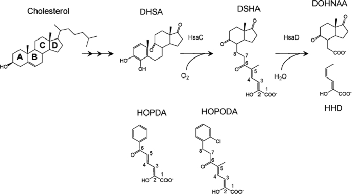
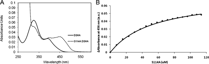
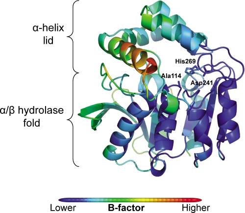
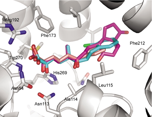
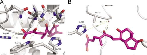
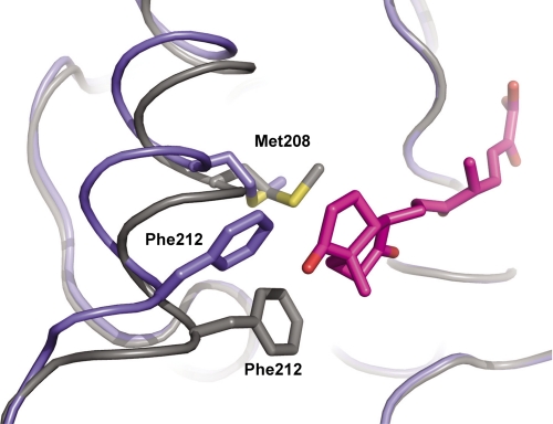
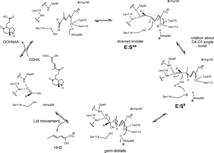
In the recently identified cholesterol catabolic pathway of Mycobacterium tuberculosis, 2-hydroxy-6-oxo-6-phenylhexa-2,4-dienoate hydrolase (HsaD) is proposed to catalyze the hydrolysis of a carbon-carbon bond in 4,5-9,10-diseco-3-hydroxy-5,9,17-tri-oxoandrosta-1(10),2-diene-4-oic acid (DSHA), the cholesterol meta-cleavage product (MCP) and has been implicated in the intracellular survival of the pathogen. Herein, purified HsaD demonstrated 4-33 times higher specificity for DSHA (k(cat)/K(m) = 3.3 +/- 0.3 x 10(4) m(-1) s(-1)) than for the biphenyl MCP 2-hydroxy-6-oxo-6-phenylhexa-2,4-dienoic acid (HOPDA) and the synthetic analogue 8-(2-chlorophenyl)-2-hydroxy-5-methyl-6-oxoocta-2,4-dienoic acid (HOPODA), respectively. The S114A variant of HsaD, in which the active site serine was substituted with alanine, was catalytically impaired and bound DSHA with a K(d) of 51 +/- 2 mum. The S114A.DSHA species absorbed maximally at 456 nm, 60 nm red-shifted versus the DSHA enolate. Crystal structures of the variant in complex with HOPDA, HOPODA, or DSHA to 1.8-1.9 Aindicate that this shift is due to the enzyme-induced strain of the enolate. These data indicate that the catalytic serine catalyzes tautomerization. A second role for this residue is suggested by a solvent molecule whose position in all structures is consistent with its activation by the serine for the nucleophilic attack of the substrate. Finally, the alpha-helical lid covering the active site displayed a ligand-dependent conformational change involving differences in side chain carbon positions of up to 6.7 A, supporting a two-conformation enzymatic mechanism. Overall, these results provide novel insights into the determinants of specificity in a mycobacterial cholesterol-degrading enzyme as well as into the mechanism of MCP hydrolases.


Figures

FIGURE 1.
FIGURE 1.
The successive reactions catalyzed by HsaC and HsaD in the catabolism of cholesterol by M. tuberculosis and alternate substrates of HsaD. DSHA is referred to as 4,5–9,10-diseco-3-hydroxy-5,9,17-trioxoandrosta-1(10),2-diene-4-oic acid in the steroid literature. However, the carbon atoms are numbered here according to its IUPAC name, 2-hydroxy-5-methyl-8-(7a-methyl-1,5-dioxooctahydro-1H-inden-4-yl)-6-oxoocta-2,4-dienoic acid, to be consistent with the numbering in HOPDA and HOPODA. In this study, DSHA, HOPDA, and HOPODA were generated by ring cleavage of DHSA, 2,3-dihydroxybiphenyl, and DHDS, respectively.
FIGURE 2.
FIGURE 2.
Characterization of the S114A·DSHA complex by UV-visible absorption spectroscopy. A, spectra of S114A·DSHA and DSHA in solution are shown by dotted and solid lines, respectively. The complex absorbs maximally at 456 nm. B, the curve represents the fit of the binding equation to ΔA456 observed upon titrating DSHA with S114A (Kd = 51 ± 2 μm). The experiments were performed using potassium phosphate (I = 0.1 m, pH 7.5) at 25 °C.
FIGURE 3.
FIGURE 3.
Secondary structure and B-factor of S114A. The secondary structure of S114A was color-coded according to Cα B-factor from blue (lowest B-factor: 6.0 Å2) to red (highest B-factor: 67.4 Å2).
FIGURE 4.
FIGURE 4.
Section of electron density map of S114A in complex with MCP. Stereo view showing 2FoFc (blue, contour level = 1 σ) and FoFc (green, contour level = 2.5σ). The electron density maps are shown for the ligand HOPDA (A), DSHA (B), and HOPODA (C). The protein structures are shown as white, whereas the ligands are shown as pink (HOPDA), purple (DSHA), or cyan (HOPODA). All of the structures are shown in ball and stick representation, with the nitrogen, oxygen, and sulfur atoms colored blue, red, and yellow, respectively.
FIGURE 5.
FIGURE 5.
Overlay of the MCP substrates HOPDA, HOPODA, and DSHA in complex with S114A. The protein (white) is shown in with the side chains involved in ligand binding shown as sticks. The ligands are represented as sticks and are colored pink (HOPDA), cyan (HOPODA), and purple (DSHA). In all structures the nitrogen, oxygen, and sulfur atoms colored blue, red, and yellow, respectively.
FIGURE 6.
FIGURE 6.
Interaction of DSHA with S114A. A, the structure of S114A (white) in complex with DSHA (purple) is shown in cartoon representations with the amino acid side chains involved in binding and DSHA displayed in stick representation. The interactions between S114A and the C-2 oxo and C-1 carboxylic acid are shown by yellow dashes, whereas the interactions between C-6 oxo and S114A are shown by red dashes. B, the structure of S114A (white) in complex with DSHA (purple). The water molecule (HOH89) is shown by a red star. All of the distances are in angstroms.
FIGURE 7.
FIGURE 7.
Conformation of the αL4 helix in the presence of the ligand DSHA. The backbone of ligand-free S114A (blue) overlaid with that of the S114A·DSHA complex (gray). The side chains Phe212, Met208, and also DSHA (purple) are shown in stick representation.
FIGURE 8.
FIGURE 8.
Proposed mechanism of HsaD. Upon binding of the MCP, His269 deprotonates the hydroxyl at C-2, generating a strained enolate intermediate, E:Sse. Protonation of C-5 by Ser114 drives tautomerization of the substrate to generate a keto intermediate, E:Sk. Ser114 is positioned to activate water for attack at the C-6 carbonyl to form the gem-diolate. The collapse of the tetrahedral intermediate releases HHD, which triggers a conformational change in the lid domain and allows subsequent release of DOHNAA.

References

    1. Dye C. (2006) Lancet 367, 938–940 - PubMed
    1. Schnappinger D., Ehrt S., Voskuil M. I., Liu Y., Mangan J. A., Monahan I. M., Dolganov G., Efron B., Butcher P. D., Nathan C., Schoolnik G. K. (2003) J. Exp. Med. 198, 693–704 - PMC - PubMed
    1. Sobel H., Plaut A. (1949) J. Bacteriol. 57, 377–382 - PMC - PubMed
    1. Van der Geize R., Yam K., Heuser T., Wilbrink M. H., Hara H., Anderton M. C., Sim E., Dijkhuizen L., Davies J. E., Mohn W. W., Eltis L. D. (2007) Proc. Natl. Acad. Sci. U.S.A. 104, 1947–1952 - PMC - PubMed
    1. Pandey A. K., Sassetti C. M. (2008) Proc. Natl. Acad. Sci. U.S.A. 105, 4376–4380 - PMC - PubMed

Publication types

MeSH terms