Review
. 2010 May;31(5):803-8.
doi: 10.3174/ajnr.A1811. Epub 2009 Oct 29.

Addictive illegal drugs: structural neuroimaging

Affiliations
Review

Addictive illegal drugs: structural neuroimaging

S Geibprasert et al. AJNR Am J Neuroradiol. 2010 May.

Abstract

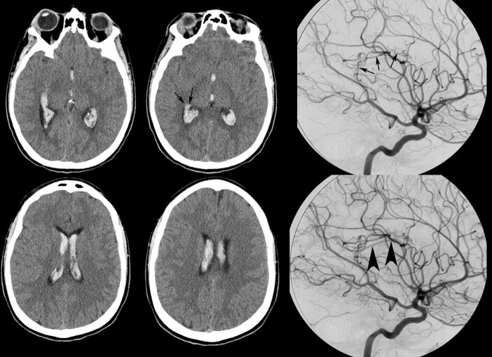
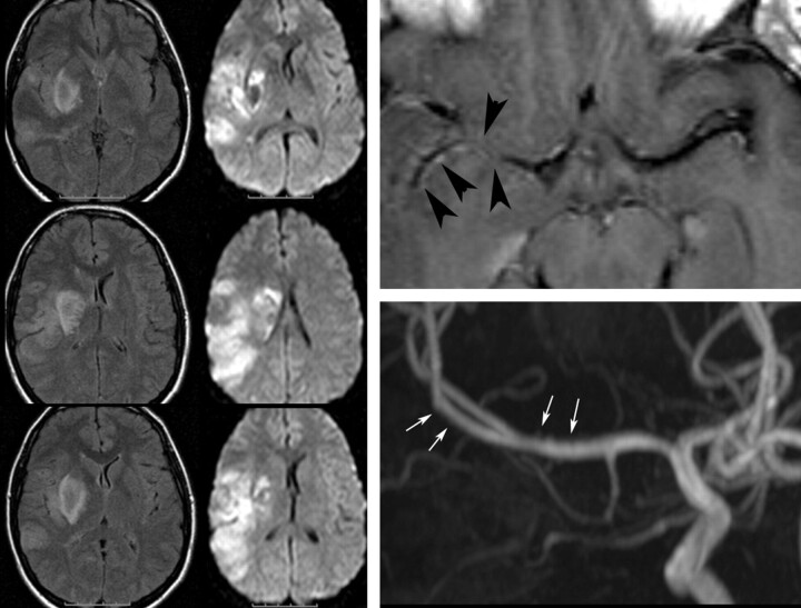
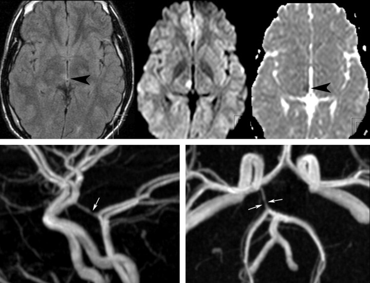
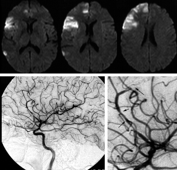
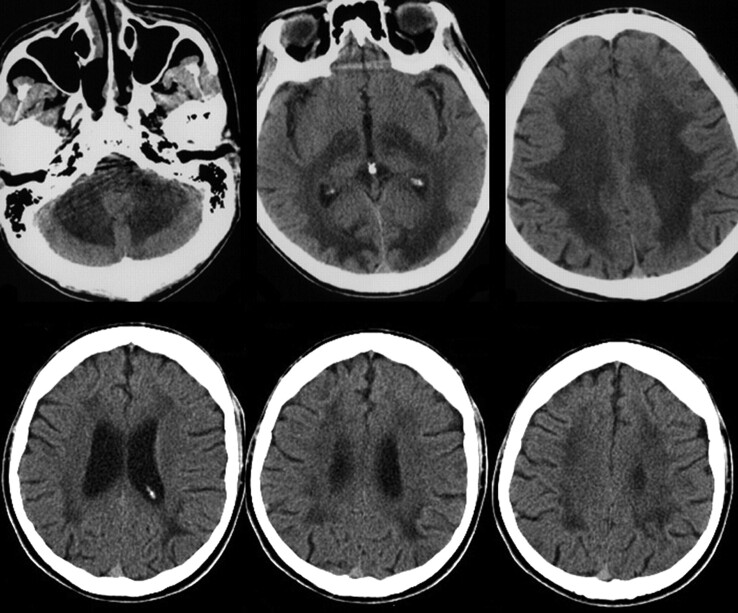
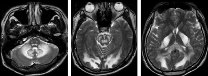
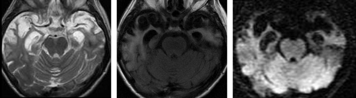
Illegal addictive drugs can lead to functional or structural impairment of the central nervous system. This review provides an overview of the structural imaging findings on CT, MR imaging, and conventional angiography related to chronic and acute abuse of the most commonly abused illegal drugs, including cannabis, organic solvents, and amphetamines and opioids and their respective derivatives. Pathomechanisms include excitotoxicity, which may lead to an acute or subacute leukoencephalopathy, and vascular complications, including vasoconstriction, vasculitis, or hypertension, which may lead to intracranial hemorrhage or ischemia. Because clinical findings alone are often nonspecific, and afflicted patients are unlikely to admit to the substance abuse, the neuroradiologist may play an important role in establishing the diagnosis and, thereby, initiating treatment.


Figures

Fig 1.
Fig 1.
A 19-year-old woman presented with acute onset of headaches to the emergency department. CT demonstrates an intraventricular hemorrhage with a potential parenchymal component close to the right ventricular wall (arrows on CT). Drug screening was positive for alcohol and cocaine. Because approximately half of the patients with intracranial hemorrhage following cocaine abuse have an underlying cause for their hemorrhage, angiography was performed, demonstrating a choroidal arteriovenous malformation fed by the posterior lateral choroidal artery (arrows on angiography) of the posterior cerebral artery, with slightly early venous filling (arrowheads), which was subsequently successfully embolized.
Fig 2.
Fig 2.
A 23-year-old woman woke up with a complete hemiplegia. Following a positive urine analysis, she admitted having taken considerable amounts of cocaine the night before. MR imaging demonstrates an MCA branch ischemia including involvement of the basal ganglia. T1-weighted postcontrast scans demonstrate abnormal vessel wall enhancement in the right MCA (arrowheads), and the time-of-flight MRA shows some mild irregularities of the distal M1, potentially testifying to acute drug-induced vasculitis as the most likely cause for the ischemia (arrows).
Fig 3.
Fig 3.
This young male patient with a subacute onset of hemisensory deficit following ingestion of an unidentified psychoactive designer drug has a small area of cytotoxic edema with fluid-attenuated inversion recovery hyperintensity and decreased apparent diffusion coefficient value in his right mediodorsal thalamus, which is classically supplied by thalamoperforators arising from the distal posterior communicating artery (arrowheads). The time-of-flight MRA at 3T demonstrates a focal narrowing of this artery at the presumed origin of the thalamoperforators supplying the infarcted region (arrows). Drug-induced vasospasm or local vasculitis is presumed to be the cause for his acute ischemia.
Fig 4.
Fig 4.
Acute areas of ischemia in both the anterior and middle cerebral artery territories involving predominantly the gray matter as a result of a diffuse vasculitis with a “pearl-and-string” sign along the anterior cerebral artery, with an acute occlusion of the pericallosal artery (arrows) and luminal irregularities in multiple different vessels in a patient with intravenous heroine abuse. Whether the described vasculitic pattern is due to vasospasm (ie, the primary effect of the opioid), immune-mediated responses against the heroin or added impurities, or a primary infectious process of the vessel wall cannot be differentiated.
Fig 5.
Fig 5.
Acute (upper row) and chronic (after 6 months) effects following heroin inhalation (chasing the dragon). In the acute phase, a diffuse edematous swelling of the white matter is present both supra- and infratentorially. On follow-up after 6 months, the patient demonstrated mental retardation and persistent psychosis. CT demonstrates bilateral degeneration of the white matter with concomitant widening of the ventricles.
Fig 6.
Fig 6.
Chronic effects of heroin inhalation (typical form). Diffuse white matter changes are present, with sparing of the dentate nuclei and the cerebellar cortex and involvement of the corticospinal tracts and medial lemnisci. Clinically, this patient presented with a progressive spastic paraparesis.
Fig 7.
Fig 7.
Chronic effects of heroin inhalation (atypical form). This patient presented with psychosis and progressive dementia 20 years after she discontinued heroin inhalation (which she performed for nearly 10 years). Although not proved, the vacuolic degeneration speaks in favor of heroin-induced leukoencephalopathy, which is predominant in the white matter of both temporal lobes.

References

    1. Fowler JS, Volkow ND, Kassed CA, et al. . Imaging the addicted human brain. Sci Pract Perspect 2007;3:4–16 - PMC - PubMed
    1. Gatley SJ, Volkow ND. Addiction and imaging of the living human brain. Drug Alcohol Depend 1998;51:97–108 - PubMed
    1. Lingford-Hughes AR, Davies SJ, McIver S, et al. . Addiction. Br Med Bull 2003;65:209–22 - PubMed
    1. Magalhaes AC. Functional magnetic resonance and spectroscopy in drug and substance abuse. Top Magn Reson Imaging 2005;16:247–51 - PubMed
    1. Mena JC, Cuellar H, Vargas D, et al. . PET and SPECT in drug and substance abuse. Top Magn Reson Imaging 2005;16:253–56 - PubMed

MeSH terms