. 2009 Nov 17;106(46):19551-6.
doi: 10.1073/pnas.0907655106. Epub 2009 Oct 29.

Evolution amplified processing with temporally dispersed slow neuronal connectivity in primates

Affiliations

Evolution amplified processing with temporally dispersed slow neuronal connectivity in primates

Roberto Caminiti et al. Proc Natl Acad Sci U S A. .

Abstract

The corpus callosum (CC) provides the main route of communication between the 2 hemispheres of the brain. In monkeys, chimpanzees, and humans, callosal axons of distinct size interconnect functionally different cortical areas. Thinner axons in the genu and in the posterior body of the CC interconnect the prefrontal and parietal areas, respectively, and thicker axons in the midbody and in the splenium interconnect primary motor, somatosensory, and visual areas. At all locations, axon diameter, and hence its conduction velocity, increases slightly in the chimpanzee compared with the macaque because of an increased number of large axons but not between the chimpanzee and man. This, together with the longer connections in larger brains, doubles the expected conduction delays between the hemispheres, from macaque to man, and amplifies their range about 3-fold. These changes can have several consequences for cortical dynamics, particularly on the cycle of interhemispheric oscillators.


Conflict of interest statement

The authors declare no conflict of interest.

Figures
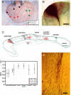
Fig. 1.
Fig. 1.
Summary of tracer studies in the macaque. Area location (in parentheses) of BDA injections in macaques CCT1–3, as determined by cytoarchitectonic criteria and according to Paxinos et al. (24). (A) Position of the injections on a lateral view of CCT2 brain; the calipers are open at 1 cm. (B) Example of BDA injection site (CCT3, near the 9/46 border); the calibration is 1 cm, and lateral is up. (C) Location of axons of different origin in cross-sections of CC rescaled and rotated to fit; the calibrations are 2 mm, anterior is to the left, and dorsal is up. Notice that the injections label discrete clusters of axons corresponding to the anteroposterior location of the injections but extending over the whole dorsal-to-ventral CC. (D) Mean diameter of axons of different origin transversally cut at the CC midline. Notice the progression of axon diameters up to, roughly, the midbody and that primary motor, somatosensory, and visual areas project thicker axons than the association areas. (E) Defasciculation of CC axons close to entering the CC explains the dorsoventral spread of axons at the CC midline, as shown in C. Calibration is 100 μm, dorsal is to the left, and the CC midline is near the bottom.
Fig. 2.
Fig. 2.
Distribution of myelinated axon diameters in different species. (Left) Photomicrographs of axons in the “motor” region of the body of the CC of macaque, chimpanzee, and human cases. Calibration is 20 μm. (Right) Histograms of the distribution of axon diameters in the 3 species. Notice the appearance of large-diameter axons in the chimpanzee and human compared with the macaque.
Fig. 3.
Fig. 3.
Distribution of conduction delays to the CC midline in different species. (Top Left) Mean conduction delays to the CC midline in each species plotted against normalized anteroposterior CC dimension and fitted with a polynomial function; “traced” refers to the BDA-traced axons and the other symbols refer to the myelinated axons. (Top Right) Mean range of conduction delays in each species. (Bottom) Histograms of the distribution of conduction velocity of myelinated axons in the “prefrontal” and “motor” sectors of the CC in the specimens of 3 species.
Fig. 4.
Fig. 4.
Interplay of interhemispheric delays regulates the cycle of cortical oscillators. (Top Left) Excitatory (E1 and E1′) and inhibitory (I2′ and I2′) Hindmarsch–Rose type neurons in a bursting mode were interconnected according to experimental findings (21). Thicker arrows indicate faster (in general) connections (21). (Top Right) Examples of bursting at 2 values of the delay of excitatory-to-inhibitory (dei) and delay of excitatory-to-excitatory (dee) connections of 20 ms. Notice that the duration of the bursts increases with dei 15 compared with dei 5. (Bottom) Relation between dei and burst length for different values of dee. Notice that increasing the range of dei values from the macaque to human amplifies the domain of interhemispheric regulation of burst length.

References

    1. Ringo JL, Doty RW, Demeter S, Simard PY. Time is of the essence: A conjecture that hemispheric specialization arises from interhemispheric conduction delay. Cereb Cortex. 1994;4:331–343. - PubMed
    1. Olivares R, Montiel J, Aboitiz F. Species differences and similarities in the fine structure of the mammalian corpus callosum. Brain Behav Evol. 2001;57:98–105. - PubMed
    1. Innocenti GM, Aggoun-Zouaoui D, Lehmann P. Cellular aspects of callosal connections and their development. Neuropsychologia. 1995;33:961–987. - PubMed
    1. Wang SS-H, et al. Functional trade-offs in white matter axonal scaling. J Neurosci. 2008;28:4047–4056. - PMC - PubMed
    1. LaMantia A-S, Rakic P. Cytological and quantitative characteristics of four cerebral commissures in the rhesus monkey. J Comp Neurol. 1990;291:520–537. - PubMed

Publication types

LinkOut - more resources