

ER stress depresses NF-kappaB activation in mesangial cells through preferential induction of C/EBP beta
- PMID: 19875812
- PMCID: PMC2799283
- DOI: 10.1681/ASN.2009040432
ER stress depresses NF-kappaB activation in mesangial cells through preferential induction of C/EBP beta
Abstract
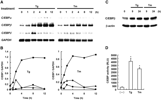
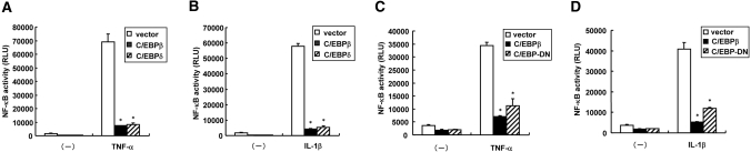
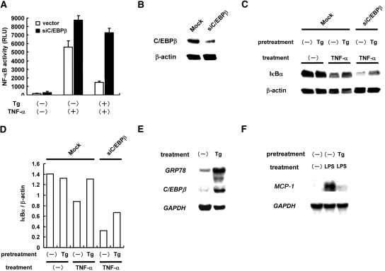
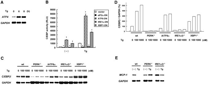
Modest induction of endoplasmic reticulum (ER) stress confers resistance to inflammation in glomeruli. Recently, we found that ER stress leads to mesangial insensitivity to cytokine-induced activation of NF-kappaB, but the underlying mechanisms are incompletely understood. ER stress can trigger expression of CCAAT/enhancer-binding proteins (C/EBPs), which interact with transcription factors including NF-kappaB. Here, we investigated a role for C/EBPs in the ER stress-induced resistance to cytokines. Mesangial cells preferentially induced C/EBPbeta after exposure to thapsigargin or tunicamycin; induction of C/EBPdelta was modest and transient, and expression of C/EBPalpha was absent. The induction of C/EBPbeta correlated with accumulation of C/EBPbeta protein and enhanced transcriptional activity of C/EBP. Overexpression of C/EBPbeta markedly suppressed TNF-alpha-induced activation of NF-kappaB, independent of its transacting potential. Knockdown of C/EBPbeta by small interfering RNA reversed the suppressive effect of ER stress on NF-kappaB. In vivo, preconditioning of mice with ER stress induced renal C/EBPbeta and suppressed NF-kappaB-dependent gene expression in response to LPS. Using dominant negative mutants and null mutants for individual branches of the unfolded protein response, we identified the RNA-dependent protein kinase-like ER kinase (PERK) and the inositol-requiring ER-to-nucleus signal kinase 1 (IRE1) pathways as the unfolded protein response responsible for ER stress-induced C/EBPbeta. These results suggest that ER stress blunts cytokine-triggered activation of NF-kappaB, in part through PERK- and IRE1-mediated preferential induction of C/EBPbeta.
Figures
Similar articles
-
Selective abrogation of BiP/GRP78 blunts activation of NF-κB through the ATF6 branch of the UPR: involvement of C/EBPβ and mTOR-dependent dephosphorylation of Akt.Mol Cell Biol. 2011 Apr;31(8):1710-8. doi: 10.1128/MCB.00939-10. Epub 2011 Feb 7. Mol Cell Biol. 2011. PMID: 21300786 Free PMC article.
-
Selective, potent blockade of the IRE1 and ATF6 pathways by 4-phenylbutyric acid analogues.Br J Pharmacol. 2013 Oct;170(4):822-34. doi: 10.1111/bph.12306. Br J Pharmacol. 2013. PMID: 23869584 Free PMC article.
-
CCAAT/enhancing binding protein beta deletion in mice attenuates inflammation, endoplasmic reticulum stress, and lipid accumulation in diet-induced nonalcoholic steatohepatitis.Hepatology. 2007 May;45(5):1108-17. doi: 10.1002/hep.21614. Hepatology. 2007. PMID: 17464987
-
Bidirectional regulation of NF-κB by reactive oxygen species: a role of unfolded protein response.Free Radic Biol Med. 2013 Dec;65:162-174. doi: 10.1016/j.freeradbiomed.2013.06.020. Epub 2013 Jun 19. Free Radic Biol Med. 2013. PMID: 23792277 Review.
-
Diabetes as a disease of endoplasmic reticulum stress.Diabetes Metab Res Rev. 2010 Nov;26(8):611-21. doi: 10.1002/dmrr.1132. Epub 2010 Oct 3. Diabetes Metab Res Rev. 2010. PMID: 20922715 Review.
Cited by
-
Endoplasmic Reticulum Stress and Its Impact on Adipogenesis: Molecular Mechanisms Implicated.Nutrients. 2023 Dec 12;15(24):5082. doi: 10.3390/nu15245082. Nutrients. 2023. PMID: 38140341 Free PMC article. Review.
-
Inhibition of interleukin-6 on matrix protein production by glomerular mesangial cells and the pathway involved.Am J Physiol Renal Physiol. 2020 Jun 1;318(6):F1478-F1488. doi: 10.1152/ajprenal.00043.2020. Epub 2020 May 11. Am J Physiol Renal Physiol. 2020. PMID: 32390515 Free PMC article.
-
Endoplasmic reticulum stress in the pathogenesis of fibrotic disease.J Clin Invest. 2018 Jan 2;128(1):64-73. doi: 10.1172/JCI93560. Epub 2018 Jan 2. J Clin Invest. 2018. PMID: 29293089 Free PMC article. Review.
-
Differential Regulation of Toll-Like Receptor-Mediated Cytokine Production by Unfolded Protein Response.Oxid Med Cell Longev. 2018 Apr 24;2018:9827312. doi: 10.1155/2018/9827312. eCollection 2018. Oxid Med Cell Longev. 2018. PMID: 29849928 Free PMC article. Review.
-
Emerging functions of the unfolded protein response in immunity.Nat Immunol. 2014 Oct;15(10):910-9. doi: 10.1038/ni.2991. Nat Immunol. 2014. PMID: 25232821 Free PMC article. Review.
References
-
- Rutkowski DT, Kaufman RJ: A trip to the ER: Coping with stress. Trends Cell Biol 14: 20–28, 2004 - PubMed
-
- Kim R, Emi M, Tanabe K, Murakami S: Role of the unfolded protein response in cell death. Apoptosis 11: 5–13, 2006 - PubMed
-
- Kitamura M: Endoplasmic reticulum stress and unfolded protein response in renal pathophysiology: Janus faces. Am J Physiol Renal Physiol 295: F323–F342, 2008 - PubMed
-
- Cybulsky AV, Takano T, Papillon J, Khadir A, Liu J, Peng H: Complement C5b-9 membrane attack complex increases expression of endoplasmic reticulum stress proteins in glomerular epithelial cells. J Biol Chem 277: 41342–41351, 2002 - PubMed
Publication types
MeSH terms
Substances
LinkOut - more resources
Full Text Sources
