. 2009 Oct;5(10):e1000643.
doi: 10.1371/journal.ppat.1000643. Epub 2009 Oct 30.

Mycobacteria exploit host hyaluronan for efficient extracellular replication

Affiliations

Mycobacteria exploit host hyaluronan for efficient extracellular replication

Yukio Hirayama et al. PLoS Pathog. 2009 Oct.

Abstract

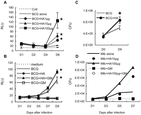
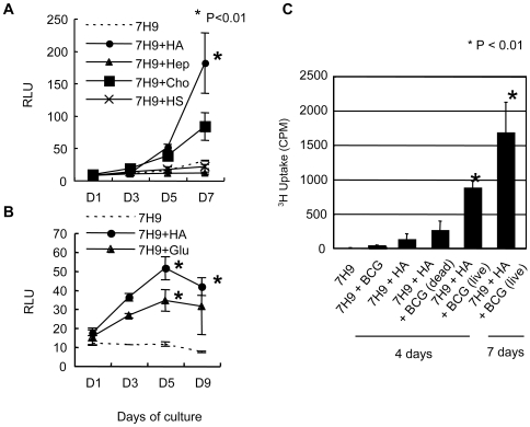
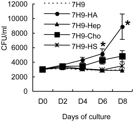
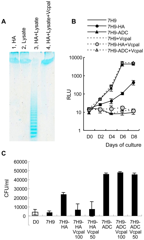
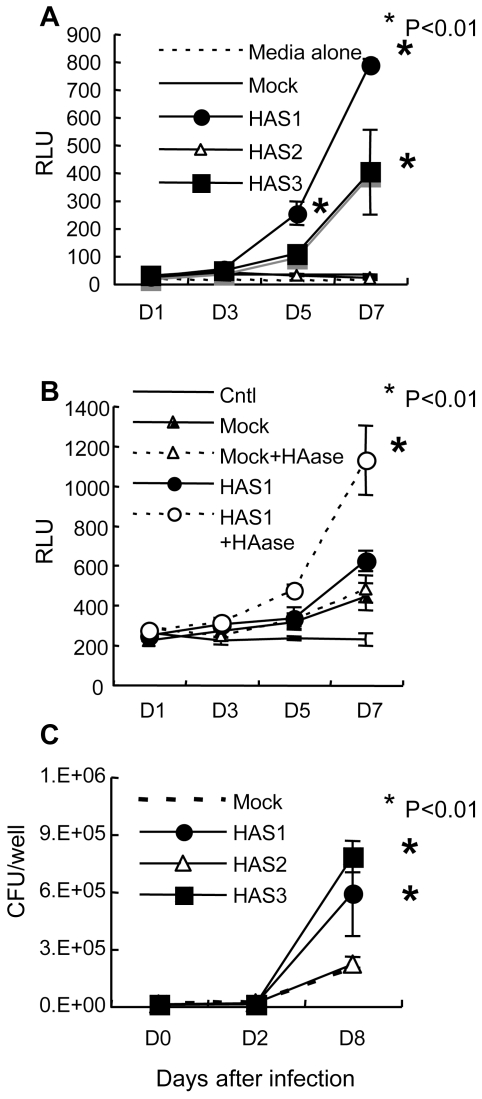
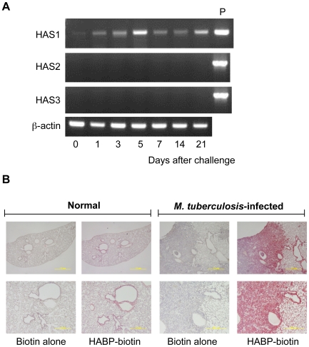
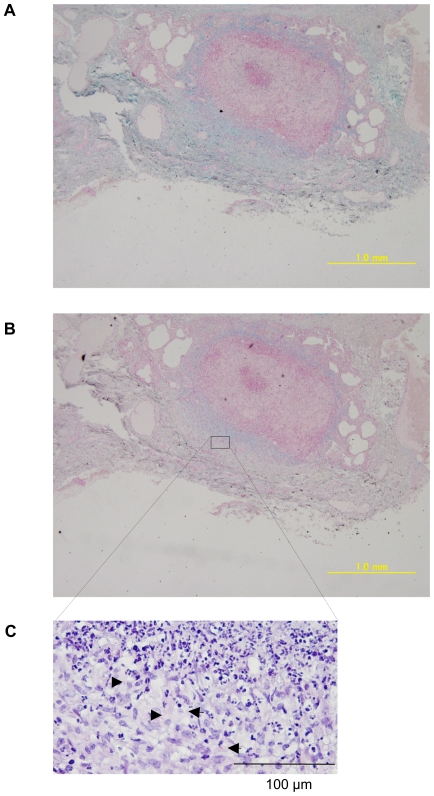
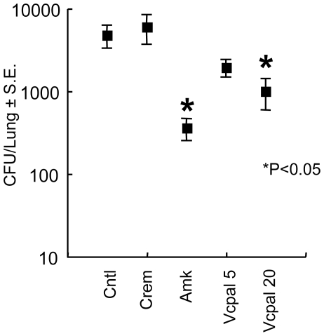
In spite of the importance of hyaluronan in host protection against infectious organisms in the alveolar spaces, its role in mycobacterial infection is unknown. In a previous study, we found that mycobacteria interact with hyaluronan on lung epithelial cells. Here, we have analyzed the role of hyaluronan after mycobacterial infection was established and found that pathogenic mycobacteria can grow by utilizing hyaluronan as a carbon source. Both mouse and human possess 3 kinds of hyaluronan synthases (HAS), designated HAS1, HAS2, and HAS3. Utilizing individual HAS-transfected cells, we show that HAS1 and HAS3 but not HAS2 support growth of mycobacteria. We found that the major hyaluronan synthase expressed in the lung is HAS1, and that its expression was increased after infection with Mycobacterium tuberculosis. Histochemical analysis demonstrated that hyaluronan profoundly accumulated in the granulomatous legion of the lungs in M. tuberculosis-infected mice and rhesus monkeys that died from tuberculosis. We detected hyaluronidase activity in the lysate of mycobacteria and showed that it was critical for hyaluronan-dependent extracellular growth. Finally, we showed that L-Ascorbic acid 6-hexadecanoate, a hyaluronidase inhibitor, suppressed growth of mycobacteria in vivo. Taken together, our data show that pathogenic mycobacteria exploit an intrinsic host-protective molecule, hyaluronan, to grow in the respiratory tract and demonstrate the potential usefulness of hyaluronidase inhibitors against mycobacterial diseases.


Conflict of interest statement

The authors have declared that no competing interests exist.

Figures

Figure 1
Figure 1. Effect of exogenously added hyaluronan on the growth of BCG and M. tuberculosis after infection of A549 cells.
(A), A549 cells were infected with BCG-Luc for 16 hours at a multiplicity of infection (MOI) of 10. After removal of non-infected bacteria, different amounts of hyaluronan (HA) were added; 0 µg/200 µl (BCG alone), 1 µg/200 µl (BCG+HA1µg), 10 µg/200 µl (BCG+HA10µg), and 100 µg/200 µl (BCG+HA100µg) before culture at 37°C under 5% CO2. Cells were lysed by adding 5% Triton X (0.5% final) at each time point (1, 2, 4, and 6 days) and bacterial growth was monitored by luciferase activity. The results are expressed as mean±the standard deviation (n = 3). Relative luciferase unit (RLU). Cntl, control without BCG-Luc infection. For statistical analysis, a two-way ANOVA with Bonferroni Post tests were used to obtain P-values for each time point, comparing the various growth conditions to the control. *P<0.01. (B), Gentamicin (GM) treatment abrogated the growth of BCG-Luc after infection of A549 cells. A549 cells were infected with BCG-Luc for 16 hours at MOI of 10. After removal of non-infected bacteria, hyaluronan was added to be 500 µg/ml for some wells (BCG+HA, BCG+HA+GM) and cultured at 37°C under 5% CO2 in the presence or absence of 10 µg/ml GM (BCG+HA+GM, BCG+GM). Growth of BCG was monitored by luciferase activity. The results are expressed as mean±the standard deviation (n = 3). RLU. Cntl, control without BCG-Luc infection. (C), The enhancing effect of hyaluronan on BCG growth was confirmed by colony forming unit (CFU). A549 cells were infected with BCG-Luc for 16 hours at MOI of 10. After removal of non-infected bacteria, BCG-Luc was grown in the presence or absence of 50 µg/ml HA. Cells were lysed at each time point and serial 10-fold dilutions were plated in duplicate on Middlebrook 7H11 agar (Difco) supplemented with oleic acid, albumin, dextrose and catalase (Difco). After incubation for 3–4 weeks at 37°C, colonies were counted and the number of CFU was calculated per well (1 ml). The results are expressed as mean±the standard deviation (n = 6). (D), A549 cells were infected with M. tuberculosis H37Rv and then different amounts of hyaluronan (HA) were added; 0 µg/200 µl (Mtb alone), 10 µg/200 µl (Mtb+HA10µg), and 100 µg/200 µl (Mtb+HA100µg). Gentamycin (50 µg/ml) was added to some wells with (Mtb+HA100 µg+GM) or without (Mtb+GM) 100 µg/200 µl hyaluronan. Cells were lysed by adding 5% Triton X (0.5% final) and the number of viable bacteria was determined by plating dilutions of the samples for CFU on 7H11-OADC agar.
Figure 2
Figure 2. Effect of hyaluronan on BCG growth in carbon-starved 7H9 media.
(A) (B), BCG-Luc was cultured in carbon-starved 7H9 media (7H9), or carbon-starved 7H9 media supplemented with 500 µg/ml of HA (7H9+HA), heparin (7H9+Hep), chondroitin sulfate C (7H9+Cho), heparan sulfate (7H9+HS), or glucose (7H9+Glu) at 37°C. Growth of BCG was monitored by luciferase activity. The results are expressed as mean±the standard deviation (n = 3). For statistical analysis, a two-way ANOVA with Bonferroni Post tests were used to obtain P-values for each time point, comparing the various growth conditions to the control. *P<0.01. (C), Uptake of 3H-hyaluronan (HA) by BCG in carbon-starved 7H9 media. Live and heat-killed BCG cells were cultured in carbon-starved 7H9 media in the presence or absence of 3H-labeled hyaluronan for 4 or 7 days. The uptake of 3H-laveled hyaluronan was measured by a gamma counter.
Figure 3
Figure 3. Effect of GAG on the growth of M. tuberculosis in carbon starved media.
M. tuberculosis H37Rv was cultured in carbon-starved 7H9 media (7H9), or carbon-starved 7H9 media supplemented with 500 µg/ml of HA (7H9+HA), heparin (7H9+Hep), chondroitin sulfate C (7H9+Cho), or heparan sulfate (7H9+HS) at 37°C. Bacterial numbers were monitored by determining CFU at each time point. The results are expressed as mean±the standard deviation (n = 3). For statistical analysis, a two-way ANOVA with Bonferroni Post tests were used to obtain P-values for each time point, comparing the various growth conditions to the control. *P<0.01.
Figure 4
Figure 4. Hyaluronidase activity in mycobacteria and the effect of hyaluronidase inhibitor on hyaluronan-dependent growth of BCG and M. tuberculosis.
(A), One mg/ml of hyaluronan and 700 µg/ml of BCG cell lysate was mixed and incubated for 3 days in the presence (HA+Lysate+Vcpal) or absence (HA+Lysate) of ascorbic palmitate (Vcpal), an inhibitor of hyaluronidase. As controls, hyaluronan alone (lane 1, HA) or BCG cell lysate alone (lane 2, Lysate) was treated in the same way. Hyaluronan was precipitated by ethanol after phenol extraction and resolved in water. Then hyaluronan was fractionated by PAGE gel electrophoresis and visualized by staining with alcian blue. (B), BCG-Luc (0.01 OD at 630 nm) was cultured in carbon-starved 7H9 media (7H9), media containing hyaluronan (500 µg/ml) as a sole carbon source (7H9-HA), or complete 7H9-ADC media (7H9-ADC) in the presence or absence of 25 µM Vcpal (+Vcpal), an inhibitor of hyaluronidase. The growth of bacteria was monitored by luciferase activity. RLU, relative luciferase unit (RLU). The results are expressed as mean±the standard deviation (n = 3). (C), The effect of Vcpal on the growth of M. tuberculosis. M. tuberculosis H37Rv was cultured in carbon starved 7H9 media (7H9), media containing 100 µg/ml hyaluronan as a sole carbon source (7H9-HA), or conventional 7H9-ADC media (7H9-ADC) with or without 50 (50) or 100 (100) µM of Vcpal for 8 days (closed bars). Bacterial number was determined by plating dilutions for CFU on 7H9-OADC agar and compared to that of Time 0 (D0, open bar).
Figure 5
Figure 5. The effect of 3 hyaluronan synthases on the growth of BCG and M. tuberculosis.
(A), Established transfectant cells (Rat 3Y1 fibroblasts) with control vector (Mock) or vector to express hyaluronan synthase 1 (HAS1), HAS2 (HAS2), or HAS3 (HAS3) were cultured in the presence of BCG-Luc or media alone. The growth of bacteria was monitored by luciferase activity. RLU, relative luciferase unit. The results are expressed as mean±the standard deviation (n = 3). For statistical analysis, a two-way ANOVA with Bonferroni Post tests were used to obtain P-values for each time point, comparing the various growth conditions to the control. *P<0.01. (B), Hyaluronidase (HAase) treatment enhances the growth of BCG after infection to HAS1-tranfected cells. After 16 hours exposure of BCG-Luc to transfected cells with control vector (Mock) or vector expressing HAS1 (HAS1), unbound bacteria were washed and cultured in the presence or absence of 2 units/ml of hyaluronidase (HAase). Bacterial growth was monitored by the luciferase activity (RLU). Cntl, HAS1-transfectant cells without infection of BCG-luc. The results are expressed as mean±the standard deviation (n = 3). For statistical analysis, a two-way ANOVA with Bonferroni Post tests were used to obtain P-values for each time point, comparing the various growth conditions to the control. *P<0.01. (C), The growth of M. tuberculosis H37Rv after infection to transfectant 3Y1 fibroblasts with control vector (Mock) or vector to express hyaluronan synthase 1 (HAS1), HAS2 (HAS2), or HAS3 (HAS3) was monitored by CFU. The results are expressed as mean±the standard deviation (n = 3). For statistical analysis, a two-way ANOVA with Bonferroni Post tests were used to obtain P-values for each time point, comparing the various growth conditions to the control. *P<0.01.
Figure 6
Figure 6. Production of hyaluronan during M. tuberculosis infection in mice.
(A), BALB/c mice were aerogenically infected with M. tuberculosis H37Rv (around 10 CFU/lung). At the indicated periods, mice were euthanized and total RNA was extracted from the lungs. Transcription of each gene encoding HAS1, HAS2, HAS3 and beta-actin was analyzed by RT-PCR. Three mice were analyzed for each time point and representative data are presented. P, positive control of PCR employing the cDNA clone of each HAS gene as a template. (B), After euthanized, lungs from uninfected mice (Normal) or mice 21 days after infection with M. tuberculosis H37Rv (M. tuberculosis infected) were removed and histological sections were made by standard methods including formalin fixation, dehydration, and embedding in paraffin. Biotinylated hyaluronan-binding protein (HABP-biotin) was used to stain the hyaluronan in the lungs. Biotin alone was used as control straining (Biotin alone). Avidin-conjugated alkaline phosphatase and chromogen as the substrate were used to generate a red reaction product. Digital images of representative sites were acquired at ×20 (upper pictures) or ×100 (lower pictures) magnification. Experiments were performed at least three times using 5 mice for each group.
Figure 7
Figure 7. Presence of hyaluronan in the lungs of rhesus monkeys that died from tuberculosis.
The lung sections were obtained from rhesus monkeys that had died of tuberculosis after challenge with 3,000 CFU/lung of M. tuberculosis H37Rv intratracheally. The sections were stained with alcian blue with (B) or without (A) pretreatment of hyaluronidase and counterstained with nuclear fast red. The section was also stained with Ziehl-Neelsen to demonstrate the presence of acid-fast bacilli (arrow heads) (C).
Figure 8
Figure 8. Vcpal suppresses the growth of mycobacteria in mouse lungs.
BALB/c mice were infected with 106 CFU of BCG (Pasteur) intravenously. One day after the challenge, mice were treated with amikacin (Amk) and Vcpal every day for 14 days. Two days after final treatment, mice were euthanized and their lungs were homogenized. Lung pastes were serially diluted and plated in duplicate on Middlebrook 7H11 OADC agars. After incubation for 3–4 weeks at 37°C, colonies were counted and the number of CFU was calculated per lung. For statistical analysis, a two-way ANOVA with Bonferroni Post tests were used to obtain P-values to determine the effect of Vcpal and amikacin on bacterial growth to the control. *P<0.05. Cntl, control mice without treatment.

References

    1. Aoki K, Matsumoto S, Hirayama Y, Wada T, Ozeki Y, et al. Extracellular mycobacterial DNA-binding protein 1 participates in Mycobacterium-lung epithelial cell interaction through hyaluronic acid. J Biol Chem. 2004;279:39798–39806. - PubMed
    1. Bermudez LE, Goodman J. Mycobacterium tuberculosis invades and replicates within type II alveolar cells. Infect Immun. 1996;64:1400–1406. - PMC - PubMed
    1. Hernandez-Pando R, Jeyanathan M, Mengistu G, Aguilar D, Orozco H, et al. Persistence of DNA from Mycobacterium tuberculosis in superficially normal lung tissue during latent infection. Lancet. 2000;356:2133–2138. - PubMed
    1. Teitelbaum R, Schubert W, Gunther L, Kress Y, Macaluso F, et al. The M cell as a portal of entry to the lung for the bacterial pathogen Mycobacterium tuberculosis. Immunity. 1999;10:641–650. - PubMed
    1. Nishimura J, Saiga H, Sato S, Okuyama M, Kayama H, et al. Potent antimycobacterial activity of mouse secretory leukocyte protease inhibitor. J Immunol. 2008;180:4032–4039. - PubMed

Publication types

MeSH terms

LinkOut - more resources