. 2009 Oct;5(10):e1000639.
doi: 10.1371/journal.ppat.1000639. Epub 2009 Oct 30.

Neutrophil extracellular traps contain calprotectin, a cytosolic protein complex involved in host defense against Candida albicans

Affiliations

Neutrophil extracellular traps contain calprotectin, a cytosolic protein complex involved in host defense against Candida albicans

Constantin F Urban et al. PLoS Pathog. 2009 Oct.

Abstract

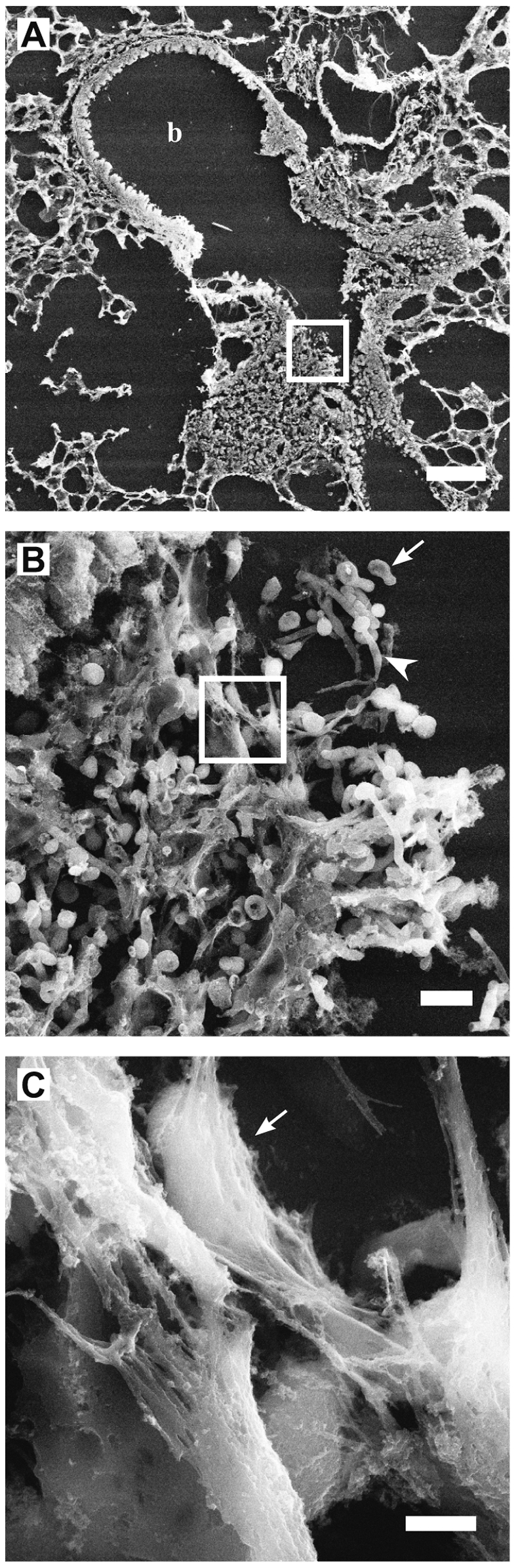
Neutrophils are the first line of defense at the site of an infection. They encounter and kill microbes intracellularly upon phagocytosis or extracellularly by degranulation of antimicrobial proteins and the release of Neutrophil Extracellular Traps (NETs). NETs were shown to ensnare and kill microbes. However, their complete protein composition and the antimicrobial mechanism are not well understood. Using a proteomic approach, we identified 24 NET-associated proteins. Quantitative analysis of these proteins and high resolution electron microscopy showed that NETs consist of modified nucleosomes and a stringent selection of other proteins. In contrast to previous results, we found several NET proteins that are cytoplasmic in unstimulated neutrophils. We demonstrated that of those proteins, the antimicrobial heterodimer calprotectin is released in NETs as the major antifungal component. Absence of calprotectin in NETs resulted in complete loss of antifungal activity in vitro. Analysis of three different Candida albicans in vivo infection models indicated that NET formation is a hitherto unrecognized route of calprotectin release. By comparing wild-type and calprotectin-deficient animals we found that calprotectin is crucial for the clearance of infection. Taken together, the present investigations confirmed the antifungal activity of calprotectin in vitro and, moreover, demonstrated that it contributes to effective host defense against C. albicans in vivo. We showed for the first time that a proportion of calprotectin is bound to NETs in vitro and in vivo.


Conflict of interest statement

The authors have declared that no competing interests exist.

Figures

Figure 1
Figure 1. Identification of NET-associated proteins.
(A) Silver stained SDS-PAGE and (B) immunoblots with samples from NET protein purification procedure. Human neutrophils were stimulated to form NETs. Supernatants from unstimulated (lane 1) and stimulated (lane 2) neutrophils; first wash (lane 3); second wash (lane 4); medium containing DNase-1 incubated with unstimulated neutrophils (lane 5); DNase-1-free medium incubated with washed NETs (lane 6); medium containing DNase-1 incubated with washed NETs (lane 7); medium containing DNase-1 incubated with washed NETs including protease inhibitor cocktail (lane 8).
Figure 2
Figure 2. Histones are altered during NET formation.
NETs from human neutrophils were washed and digested with DNase-1. (A) The NET-fraction (N) and the remaining pellet after DNase-1 digest (P) were analyzed by immunoblotting at the indicated time points. Unstimulated neutrophils served as controls. All core histones have a reduced molecular mass (2–5 kDa less) in NETs compared to the pellet fraction and the unstimulated control. A representative experiment out of three in total is shown. (B) High-resolution SEM analysis of NETs which consist of smooth fibers (white box) and globular domains (diameter 25–50 nm, arrows), scale bar = 100 nm. (C) High-resolution FESEM analysis of smooth stretch of a singular NET-fiber. Signal intensities were profiled vertically and horizontally showing similar diameters to nucleosomes (depicted as cartoon structure models taken from , with approximate horizontal and vertical diameters of 5 nm and 10 nm, respectively). One experiment out of two is shown.
Figure 3
Figure 3. Neutrophils release calprotectin by forming NETs.
(A–F) Confocal images of human neutrophils without stimulation (A), after 0.5 h (B), 1 h (C), 2 h (D), 3 h (E) and 4 h (F) after activation. Samples were stained with antibodies specific for the calprotectin heteroduplex (red) and for MPO (green). DNA was stained with DRAQ5 (blue). Calprotectin localizes to the cytoplasm and partially to the nucleus (A, arrow). After stimulation for 0.5 h (B) the neutrophils flattened and formed numerous vacuoles. This reveals a granular staining for MPO and a more dispersed cytoplasmic staining for calprotectin. After stimulation for 1 h (C) the neutrophils round up slightly. The MPO and calprotectin stain partially overlap in the cytoplasm. After stimulation for 2 h (D), calprotectin, MPO and nuclear DNA start to colocalize in the decondensed nucleus (purple). After 3 h (E) and more so after 4 h (F) of stimulation, the cell membrane ruptures and calprotectin is released in NETs colocalizing with MPO and DNA. Scale bar = 10 µm; one experiment out of two is shown. (G–I) Subunits of calprotectin S100A8 and S100A9 are released after cell death during NET formation and not by degranulation. NET formation was induced with PMA and degranulation using formyl-met-leu-phe (f-MLP). (G) Neutrophil death was monitored by quantification of LDH activity in supernatants calculated as means±s.d. (n = 3). (H) Release of S100A8, S100A9, lactotransferrin (LTF) and myeloperoxidase (MPO) were analyzed by immunoblotting. one experiment out of two is shown. (I) Quantification of immunoblots using 2D densitometry analyzing S100A9 protein preparations from supernatants (lane 1), MNase-digested NETs (lane 2) and cell remnants indigestible for MNase (lane 3). Values were calculated as means±s.d. (n = 3) from one experiment out of two.
Figure 4
Figure 4. Calprotectin is a major antifungal component in NETs.
Human neutrophils were induced to make NETs, washed and infected with C. albicans (MOI 0.01) and incubated overnight at 30°C (yeast-form growth). Antifungal activity was determined by counting CFU. Increasing concentrations of ZnSO4 (A) and of MnCl2 (B) abolished the antifungal activity of human NETs. Shown are means±s.d. (n = 3) from one representative experiment out of three. (C) Purified human NET proteins were concentrated and depleted of calprotectin using immobilized anti-S100A8 and anti-S100A9 combined. Controls were incubated with mouse IgG1 isotype matched antibodies. C. albicans was incubated with these extracts overnight at 37°C (hyphal growth) and CFU were determined. Shown are means±s.d. (n = 3) from one experiment out of two. The inset shows an immunoblot confirmation of the depletion. The blots were probed for S100A9 and lactotransferrin (LTF). The lanes are arranged in the same order as indicated for the antifungal assay below the graph. (D) Mouse neutrophils isolated either from wild-type or calprotectin-deficient mice were induced to make NETs and then infected with C. albicans (MOI 0.02) incubated overnight at 37°C (hyphal growth) and CFU counts were determined; n. d. = no detectable CFU. Shown are means±s.d. (n = 3) from one experiment out of two. (E) Co-precipitation assays of indicated fungi incubated with NET-bound calprotectin (produced by MNase treatment) or soluble calprotectin. Microbes were pelleted, supernatants removed and washed pellets were analyzed using immunoblots against S100A9. Shown are means±s.d. (n = 3) of one representative experiment out of three.
Figure 5
Figure 5. Calprotectin is required for antifungal immunity.
(A–D) Subcutaneous abscesses induced with C. albicans of representative calprotectin -deficient (a and b) and wild-type (c and d) mice at days 2, 4, 6 and 8 post infection (p.i.). (E) the area of the abscess lesions of calprotectin-deficient animals compared to wild type was monitored over 8 days p.i. (n = 10); abscesses of calprotectin-deficient animals are significantly larger at days 2 (P = 0.0007), 4 (P = 0.0012), 6 (P = 0.0003) but not at day 8. (F) calprotectin-deficient mice are more susceptible to intranasal challenge with C. albicans than wild-type mice (n = 10, P = 0.0001). (G) Fungal load in the lungs after intranasal challenge with C. albicans is significantly higher in calprotectin-deficient compared to wild-type mice (n = 11; P = 0.048). (H) Calprotectin-deficient mice are more susceptible to intravenous challenge with C. albicans than wild-type mice (n = 10, P = 0.0159).
Figure 6
Figure 6. Calprotectin is present in C. albicans induced NETs in vivo.
Wild-type mice were challenged subcutaneously (A–F) or intranasally (G–L). (A,B, G and H) Hematoxylin & Eosin (H & E) stainings of sections in areas with strong neutrophil infiltration show extracellular DNA (hematoxylin positive) representing NETs and indicated with arrows, scale bars 50 µm in (A,G) and 20 µm in (B,H). Confocal images of indirect immunofluorescence from sections of abscesses 6 days after subcutaneous challenge (C–F) and lungs 24 h after intranasal challenge (I–L) stained with primary antibodies against S100A9 (red), MPO (green) and histone (blue). NETs are web-like and diffuse areas where all signals superimpose indicated by white arrows. Scale bars = 20 µm.
Figure 7
Figure 7. Fine structure of C. albicans induced NETs in pulmonary infection.
(A–C) SEM analysis of sections from C. albicans infected mouse lungs 24 h after intranasal challenge. (A) Image shows a bronchiole colonized with C. albicans and infiltrated by host immune cells, b = bronchiole. (B) High resolution image of boxed area in (n) shows respiratory epithelium of the bronchiole colonized with C. albicans yeast-form (arrow) and hyphae (arrowhead). (C) High resolution image of boxed area in (O) showing NETs covering fungal surfaces (arrow). Scale bars in (N) = 100 µm, in (O) = 10 µm and in (P) = 2 µm.

References

    1. Geiszt M, Kapus A, Ligeti E. Chronic granulomatous disease: more than the lack of superoxide? J Leukoc Biol. 2001;69:191–196. - PubMed
    1. Nathan C. Neutrophils and immunity: challenges and opportunities. Nat Rev Immunol. 2006;6:173–182. - PubMed
    1. Brinkmann V, Reichard U, Goosmann C, Fauler B, Uhlemann Y, et al. Neutrophil extracellular traps kill bacteria. Science. 2004;303:1532–1535. - PubMed
    1. von Kockritz-Blickwede M, Goldmann O, Thulin P, Heinemann K, Norrby-Teglund A, et al. Phagocytosis-independent antimicrobial activity of mast cells by means of extracellular trap formation. Blood. 2008;111:3070–3080. - PubMed
    1. Yousefi S, Gold JA, Andina N, Lee JJ, Kelly AM, et al. Catapult-like release of mitochondrial DNA by eosinophils contributes to antibacterial defense. Nat Med. 2008;14:949–953. - PubMed

Publication types

MeSH terms